Glaucoma Care
Visionary Eye Doctors provides advanced treatment options to effectively manage glaucoma and prevent severe damage to the optic nerve.
Early detection through regular and complete eye exams is the key to protecting your vision from damage caused by glaucoma. A complete eye exam includes various tests to accurately diagnose glaucoma. It is important to have your eyes examined regularly as for most people there are usually few or no symptoms of glaucoma. At Visionary Eye Doctors, our physician Thandeka Myeni, MD is a board-certified ophthalmologist and is fellowship-trained in glaucoma. To learn more about Dr. Myeni, click here. Glaucoma is known as the silent thief of sight. Request to schedule a comprehensive eye exam appointment with Dr. Myeni here.
The first sign of glaucoma is often the loss of peripheral, or side vision, which can go unnoticed until late in the disease. Detecting glaucoma early is one reason you should have a complete exam with an eye specialist every one to two years. To make the diagnosis of glaucoma, we take into consideration a number of factors, such as the appearance of your optic nerves and your eye pressures; the results of a number of diagnostic tests such as corneal thickness, nerve fiber thickness, and visual field testing; and risk factors for glaucoma such as your age, family history of glaucoma, race, and your general health. If glaucoma has been detected, it’s best to start your glaucoma treatment as soon as possible. We offer trusted glaucoma treatments in the Washington, DC, DC area.
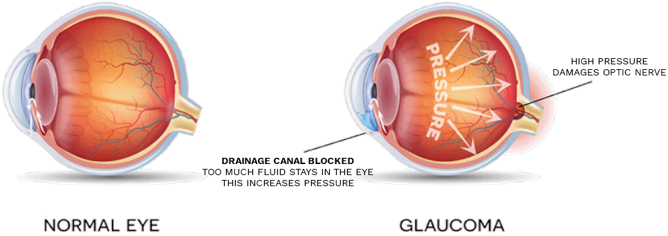
Open-angle glaucoma – Also called wide-angle glaucoma, this is the most common type of glaucoma. The structures of the eye appear normal, but fluid in the eye does not flow properly through the drain of the eye, called the trabecular meshwork.
Angle-closure glaucoma – Also called acute or chronic angle-closure or narrow-angle glaucoma, this type of glaucoma is less common but can cause a sudden build-up of pressure in the eye. Drainage may be poor because the angle between the iris and the cornea (where a drainage channel for the eye is located) is too narrow.
At Visionary Eye Doctors, we offer innovative technology for early detection of glaucoma, such as visual field testing, visual evoked potential (VEP), and electroretinography (ERG) tests.
The term visual field refers to the area of a scene you can see with your eyes fixed on one location. For example, while watching a stop light, you may notice a car pulling up beside you in the next lane. Your peripheral vision enables you to see the movement of the car. The visual field is quite extensive when both eyes are working properly but with glaucoma, the field of view is reduced.
The OCT has the ability to capture images of eye tissue at a near individual cell level of resolution. In addition, it can show each layer of tissue in the eye allowing our glaucoma eye doctors to pinpoint the exact location of tissue damage. This sophisticated piece of equipment aids in the diagnosis and management of many ocular conditions, including glaucoma and macular degeneration. OCT is a safe, painless and quick procedure that does not require dilation of the pupils. A laser scans the retina, optic nerve, cornea, or anterior chamber in just a matter of seconds to provide our glaucoma doctors with detailed information regarding the microscopic details of the eye. Anyone who has glaucoma or is a glaucoma suspect would benefit from undergoing this diagnostic test.
Pachmymetry is the measurement of the thickness of the cornea, the clear window on the front of the eye.
When the cornea is thinner than average, patients may have an increased risk of developing glaucoma. Pachymetry should be performed at least once in a glaucoma patient’s adult life if eye pressure seems too low to explain glaucomatous optic nerve damage or if the eye pressure is higher than one would expect.
As advances in technology continue to change the field of eye care, Visionary Eye Doctors is proud to offer the very latest in eye disease management. To discover vision-related diseases in their earliest stages, we use the Diopsys® NOVA Vision Testing System. Through this state-of-the-art advancement in electrophysiological technology, we can effectively detect, diagnose, and monitor progress of treatment for conditions like glaucoma and macular degeneration at their earliest stages.
Our team makes use of both the VEP (Visual Evoked Potential) and ERG (Pattern Electroretinography) protocols to conduct objective, office-based vision testing. Before the process can begin, sensors are placed on the patient’s head similar to an electrocardiogram or EKG. With such a streamlined process, the Diopsys® NOVA technology provides the following advantages:
The VEP and ERG tests each measure a specific area of the eye to thoroughly assess the function of the visual system.
The Diopsys® NOVA-VEP measures the activity of the entire visual pathway. Results from this test can determine how well the entire visual system is functioning. This test can be performed on patients as early as six months or older to detect vision or optic nerve disorders. The VEP can also be used for patients who have developed neuro-visual problems following a traumatic brain injury or stroke. It can help determine if the optic nerve function is healthy or abnormal, as seen in multiple sclerosis and glaucoma.
With the Diopsys® NOVA-ERG system, our team can determine the patient’s retinal function, which allows them to identify potential disorders. When used along with other diagnostic tests, like the VEP, this can help differentiate between a retinal and optic nerve disorder. The ERG can help detect these signs at a cellular level for earlier detection and a more timely treatment.
Considering the fact that visual diseases may develop differently, both the VEP and ERG tests are customized to the patient to fully understand their visual health. The entire process takes around 10 minutes, after which time we have instant access to the test results. The Diopsys® NOVA testing systems have proven to be an invaluable addition to our practice.
With Visionary Eye Doctors, you have many options for glaucoma treatment in the Washington, DC, DC area. Prescription Eye Drops for glaucoma help maintain the pressure in your eye at a healthy level and are an important part of the treatment routine for many people. It is important to notify your doctor of any other medications you may be taking (including over-the-counter items like vitamins, aspirin, and herbal supplements) and about any allergies you may have.
Treatment requires monitoring eye pressure and measuring changes in the peripheral visual field. Controlling glaucoma usually means maintaining eye pressure at an acceptable level when there has been little or no loss of the visual field.
We are one of the few centers in the Washington, DC, DC metro area to offer our Glaucoma patients the Icare.
The Icare home tonometer is one of the first methods for 24HR monitoring outside normal clinic hours which may help identify IOP variations and spikes.
Self-tonometry with the Icare® HOME is designed to gather additional intraocular pressure (IOP) information by the patient. The device records IOP, date, time, eye and a quality score which is not displayed to the patient. The data is retrieved by a health care provider via Icare® LINK software.
Some of the most common procedures we offer to treat glaucoma include selective laser trabeculoplasty (SLT), laser peripheral iridotomy (LPI), trabeculectomy or shunt placement.
The treatment for glaucoma has come a long way. Today’s advanced technologies and procedures make it possible to diagnose and successfully manage this progressive disease. And while the overall goal of treatment has always remained the same — reducing intraocular pressure (IOP) and glaucomatous progression — Micro-Invasive Glaucoma Surgery (MIGS) is quickly becoming a widely accepted approach for treating mild-to-moderate glaucoma.
Glaucoma is primarily managed with prescription eye drops. If a patient’s glaucoma progresses or the patient stops responding to the eye drops, a surgical solution is offered. These surgical solutions are often highly invasive, require long recovery times, and could result in long-term complications.
As technology has progressed, less invasive techniques have emerged that have improved the safety profile for glaucoma surgery. This opened more surgical options for patients interested in effective glaucoma management which does not rely solely on the continuous use of prescription medication.
With the FDA approval of the iStent®, Micro-Invasive Glaucoma Surgery (MIGS) became a preferred approach to glaucoma management for many eye care professionals and their patients with mild-to-moderate glaucoma.
Now, with the FDA approval of iStent inject® – the smallest medical device known to be implanted in the human body—is made of two tiny stents that are designed to restore your eye’s natural ability to drain fluid out of the eye to reduce glaucoma pressure. Designed for cataract patients with glaucoma, iStent inject® is implanted at the time of cataract surgery. You won’t see or feel the iStent inject® stents after they are inserted, but they are designed to effectively manage your eye pressure.
Similar to cardiac stents used in patients with heart problems, the iStent opens pressure drainage channels in the eye, permanently treating glaucoma. To help control the increased pressure in your eye associated with glaucoma, iStent inject® creates two bypasses, or openings, between the front part of your eye and its natural drainage pathway to increase the flow of fluid. By creating the permanent bypasses through the primary blockage site (trabecular meshwork), iStent inject® is designed to:
• Improve your eye’s natural outflow to safely lower intraocular pressure
• Work continuously to improve the natural flow of fluid in your eyes
If you or a family member has glaucoma, please contact us today for a consutation to discuss whether iStent or other treatments for glaucoma are an option for you. You may fill out our web form, or if you prefer, call 301.896.0890 to schedule a consultation.
Selective laser trabeculoplasty (SLT) is a glaucoma laser procedure used to lower the pressure in the eye. SLT uses short pulses of low energy laser to target specific cells in the drainage system of the eye to stimulate improved function. Ultimately, the SLT procedure can slow or stop the risk of disease progression. The SLT technique is very gentle, and studies show that it does not damage the tissue of the drainage system. The treatment takes only a few minutes to apply, and there is typically no discomfort felt by the patient. Following the laser treatment, the patient usually continues any existing glaucoma drops.
Laser Peripheral Iridotomy (LPI)
If you’d like more information about glaucoma, or to schedule a comprehensive eye exam to check for glaucoma, please request to schedule an appointment today to receive the best in eye health care.