Image of a young man getting his eyes examined by a female optometrist

Signs of Cataracts: What to Look For and When to Seek Help

Quick Take: Cataracts can cause:

    • Cloudy vision
    • Increased light sensitivity
    • Faded colors
    • Problems seeing at night

These cataract symptoms often develop slowly, and recognizing them early can help you seek appropriate treatment before your daily activities are affected.

Cataracts are one of the most common eye conditions, especially among people over 60. They occur when the eye’s natural lens becomes cloudy, similar to looking through a foggy window. While age-related cataracts develop gradually, knowing the early signs helps you maintain clear vision and quality of life.

In this blog, you’ll learn:

  • What cataracts are and why they form
  • Common signs like cloudy vision and light sensitivity
  • When to see an eye doctor
  • Treatment options, including cataract surgery
  • How to protect your vision over time

What Are Cataracts?

Cataracts form when proteins in the eye’s lens clump together, creating a clouded lens that scatters light and reduces visual clarity. This buildup leads to vision problems like blurry vision, glare, and difficulty reading or driving.

Infographic showing a diagram of two eyes: one normal and one with cataracts.

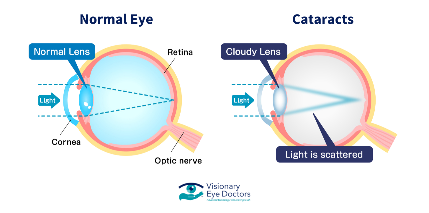
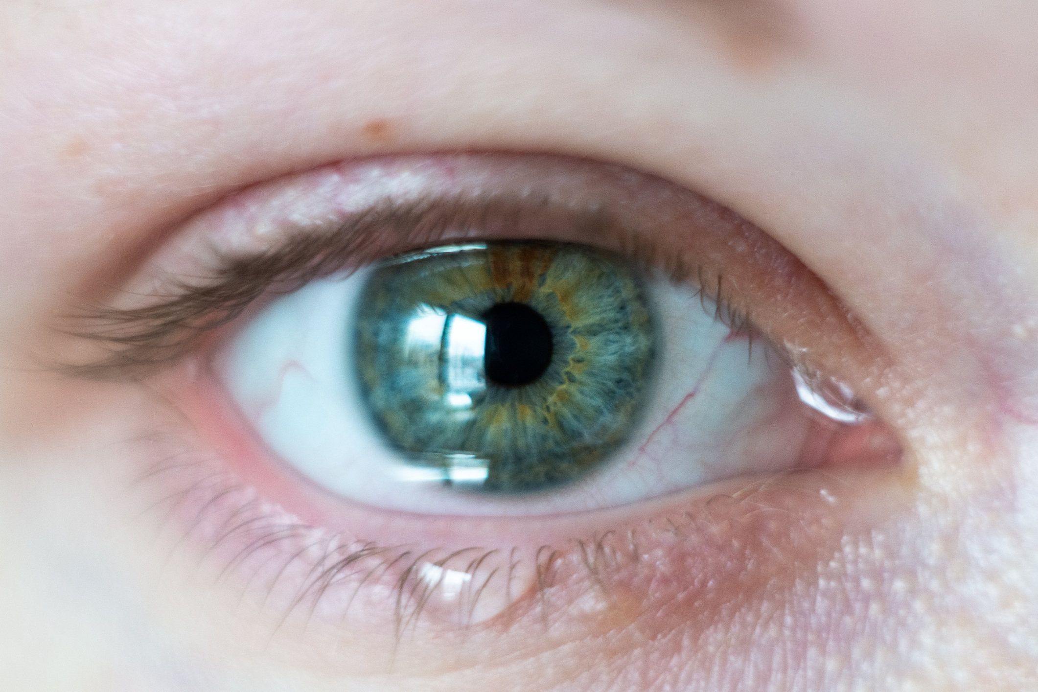
The lens sits behind your iris (the colored part of your eye) and works much like a camera lens, focusing light onto the retina at the back of your eye. A clear lens allows for sharp images on your retina, but when a cataract clouds the lens, your vision becomes blurred, similar to looking through a dirty windshield or a fogged-up window.

Types of Cataracts

There are several types of cataracts, each affecting different parts of the lens:

  • Nuclear cataract: Forms in the center of the lens and is most commonly related to aging. These cataracts often temporarily improve near vision before overall vision deteriorates.
  • Cortical cataract: Starts at the edges of the lens and gradually moves inward. These appear as wedge-shaped cloudy areas that extend from the outside edge toward the center, often resembling spokes on a wheel.
  • Posterior subcapsular cataract: Develops at the back of the lens, often leading to difficulty with bright light and night vision. These tend to progress faster than other types and frequently affect people with diabetes or those taking steroid medications.
  • Congenital cataract: Present at birth or during childhood (also known as pediatric cataracts). These can be hereditary or result from an infection during pregnancy.

What Causes Cataracts?

Cataracts can result from a range of factors, including:

  • Age, especially after 60
  • Medical conditions like diabetes or macular degeneration
  • Long-term exposure to bright sunlight or toxic substances
  • Smoking or alcohol use
  • Long-term steroid use
  • Previous eye injuries, retinal detachment, or eye surgery
  • Family history of cataracts

While aging is the primary cause, research suggests that oxidative stress plays a significant role in cataract development. This happens when unstable molecules (free radicals) attack and harm the proteins in the eye’s lens. This damage accumulates over a lifetime, which explains why cataracts become more common as we age.

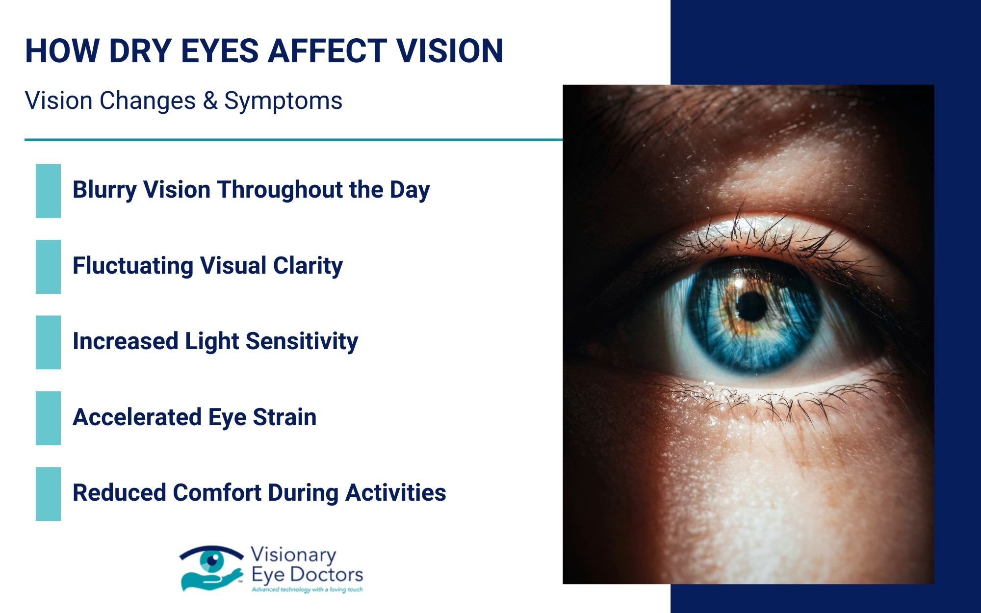
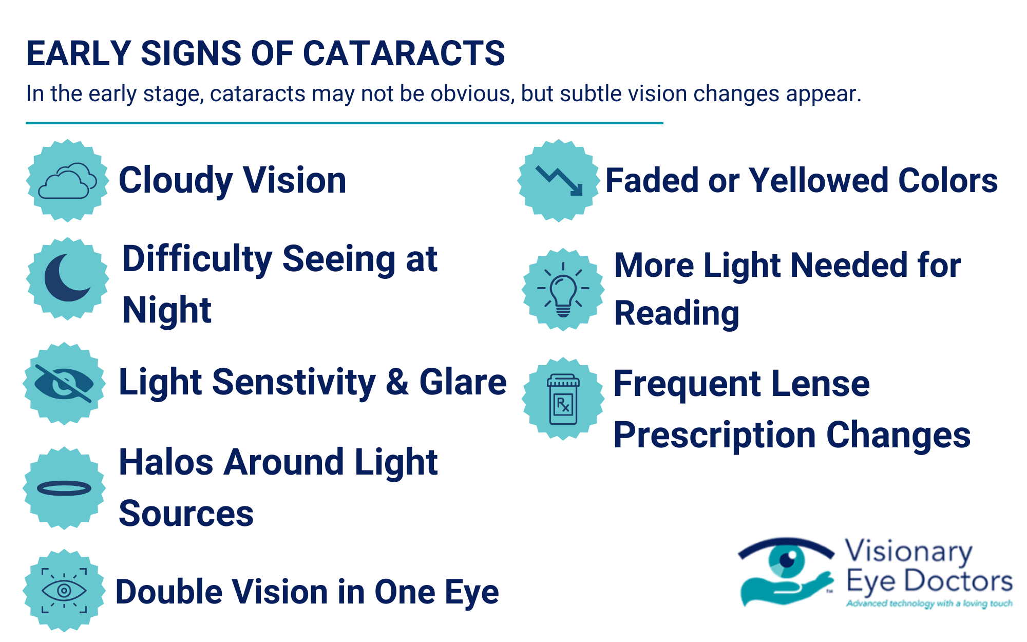
Early Signs of Cataracts

In the early stages, cataracts may not be obvious, but subtle vision changes often appear.

Common early symptoms include:

  • Cloudy vision or a sense that things look dim or hazy
  • Difficulty seeing at night or in low-light settings
  • Sensitivity to bright light and glare from headlights or sunlight
  • Seeing halos around light sources
  • Double vision in one eye
  • Colors appearing faded or yellowish
  • Needing more light to read or complete close-up tasks
  • Frequent changes to your lens prescription or contact lenses

These symptoms can come on slowly, which is why regular eye exams are essential.

Infographic depicting the early signs of cataracts including cloudy vision and light sensitivity.

Many people describe early cataract symptoms as similar to looking through a dirty car windshield or a piece of wax paper. You might find yourself cleaning your glasses frequently, thinking they’re dirty, when the problem is actually with your eye’s lens.

When Cataracts Get Worse

As the cataract progresses, the symptoms become more disruptive to your daily activities. You may notice:

  • Increasingly blurry or cloudy vision
  • Trouble distinguishing colors, especially blues and purples
  • Heightened light sensitivity and glare from bright sunlight
  • A white or yellow tint in the pupil (a possible sign of a mature cataract)
  • Temporary improvement in near vision (“second sight”), followed by worsening overall clarity

These are signs of an advanced cataract, and you should consult an eye doctor promptly.

Infographic depicting advanced cataracts symptoms, including vision changes and lens discoloration.

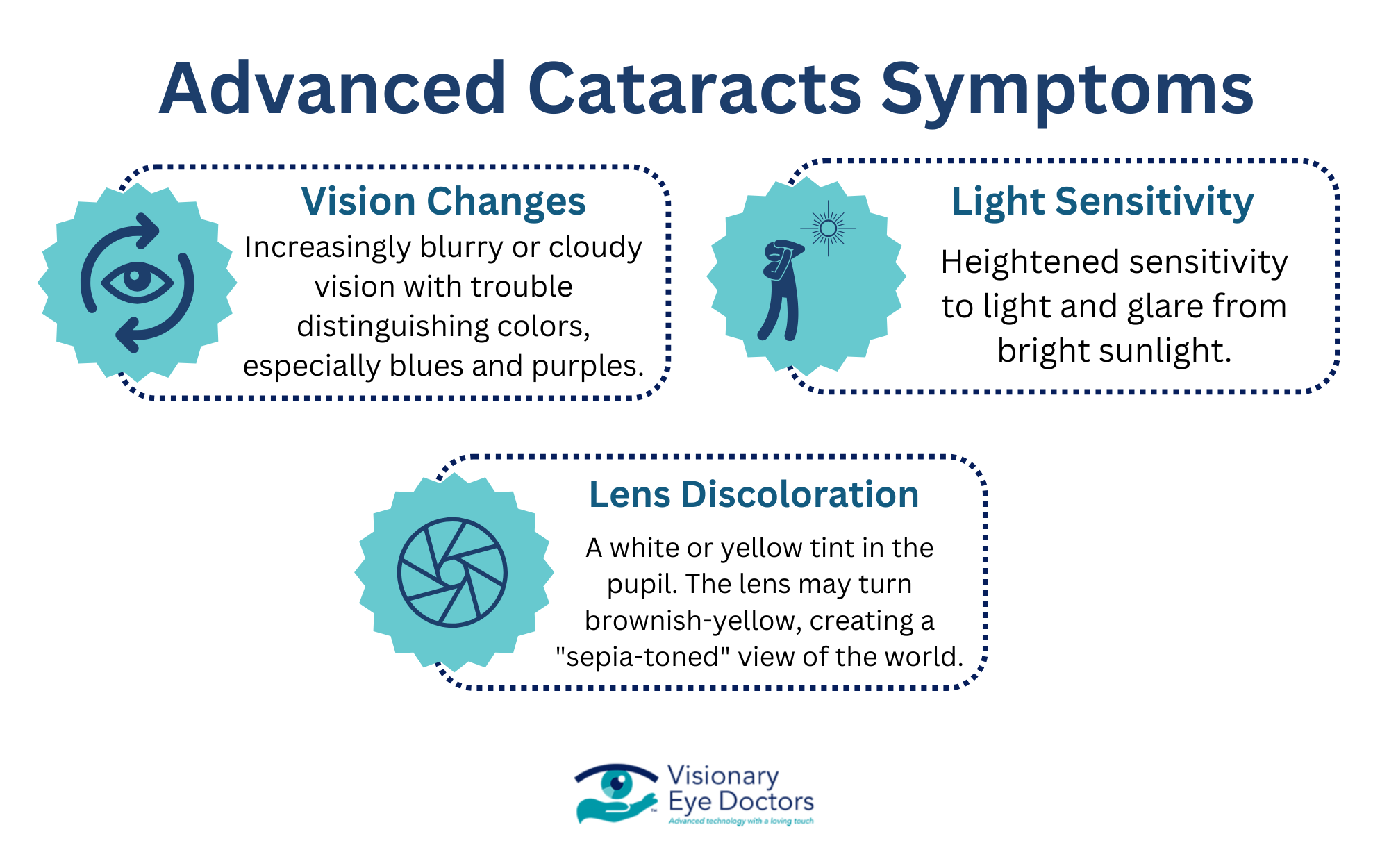
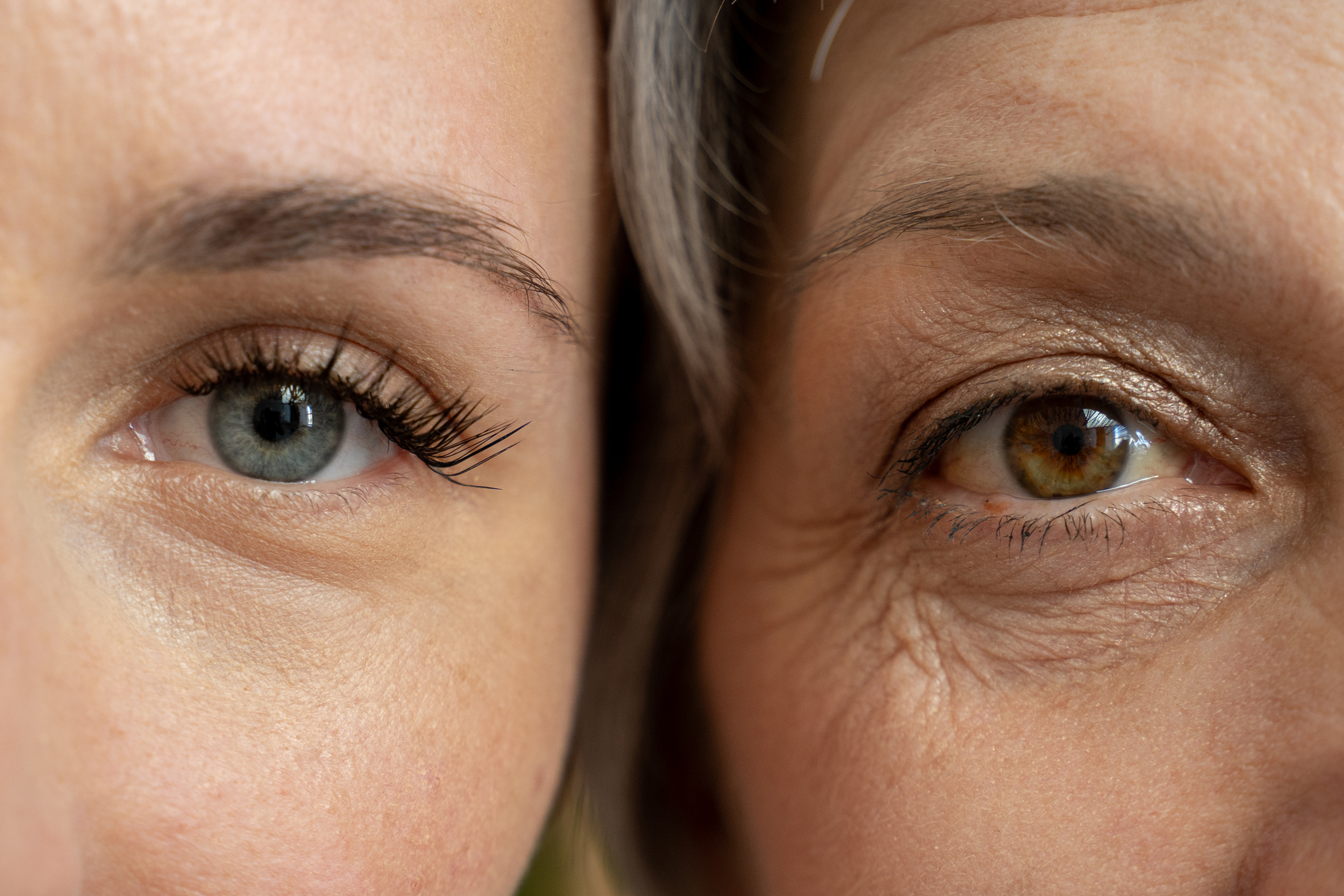
As cataracts mature, the lens may turn a brownish-yellow color, distorting your color perception. Many patients describe this as viewing the world through a sepia-toned filter. This color shift can make it difficult to distinguish between certain hues, particularly in the blue-purple spectrum.

How Cataracts Impact Daily Life

Cataracts can affect your ability to function comfortably at home, work, and on the road.

You might experience:

  • Driving difficulties – especially with night vision or glare from headlights
  • Reading problems – needing more light or magnification to see small print
  • Trouble recognizing faces from a distance
  • Difficulty with tasks that require visual detail, like sewing or cooking

If vision loss starts to interfere with normal routines, it’s time to consider your treatment options.

The impact of cataracts often extends beyond just vision problems. Many people experience increased anxiety, particularly around driving at night or in unfamiliar places. Social isolation can occur when individuals begin avoiding activities they once enjoyed because of vision difficulties. Studies show that untreated cataracts significantly increase the risk of falls and accidents, especially in older adults.

When to See an Eye Doctor

You should schedule an eye exam if:

  • You notice vision changes that make it harder to perform everyday tasks
  • Colors appear dull or yellowed
  • You’ve had multiple updates to your glasses or contact lenses
  • Glare or halos interfere with driving or reading
  • Any of the above symptoms of cataracts are affecting your comfort or independence

Recommended exam schedule:

Age Recommended Exam Frequency
40-54 Every 2-4 years
55-64 Every 1-3 years
65+ Every 1-2 years
*Patients with underlying conditions (diabetes, family history)* Annually

Diagnosing Cataracts

To confirm a cataract diagnosis, your eye doctor may:

  • Perform a visual acuity test
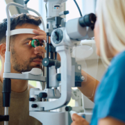
  • Use a slit-lamp microscope to examine the clouded lens
  • Dilate your pupils to inspect the retina
  • Test how you respond to glare and how well you perceive contrast
  • Evaluate color vision and depth perception

These evaluations help determine the best course of treatment, including whether surgery is needed.

During a comprehensive eye exam, your doctor will also check for other eye conditions that may coexist with cataracts, such as glaucoma or age-related macular degeneration. This thorough approach ensures that all potential vision threats are identified and addressed.

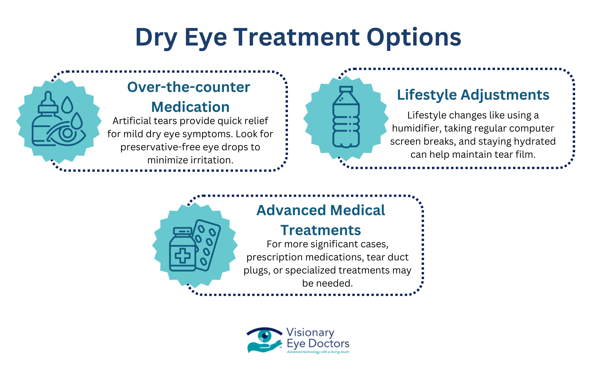
Cataract Treatment Options

Non-Surgical Management

In mild cases, especially during the early stages, symptoms can often be managed through:

  • Updated lens prescriptions
  • Anti-glare coatings on glasses
  • Brighter lighting at home
  • Use of magnifiers or visual aids
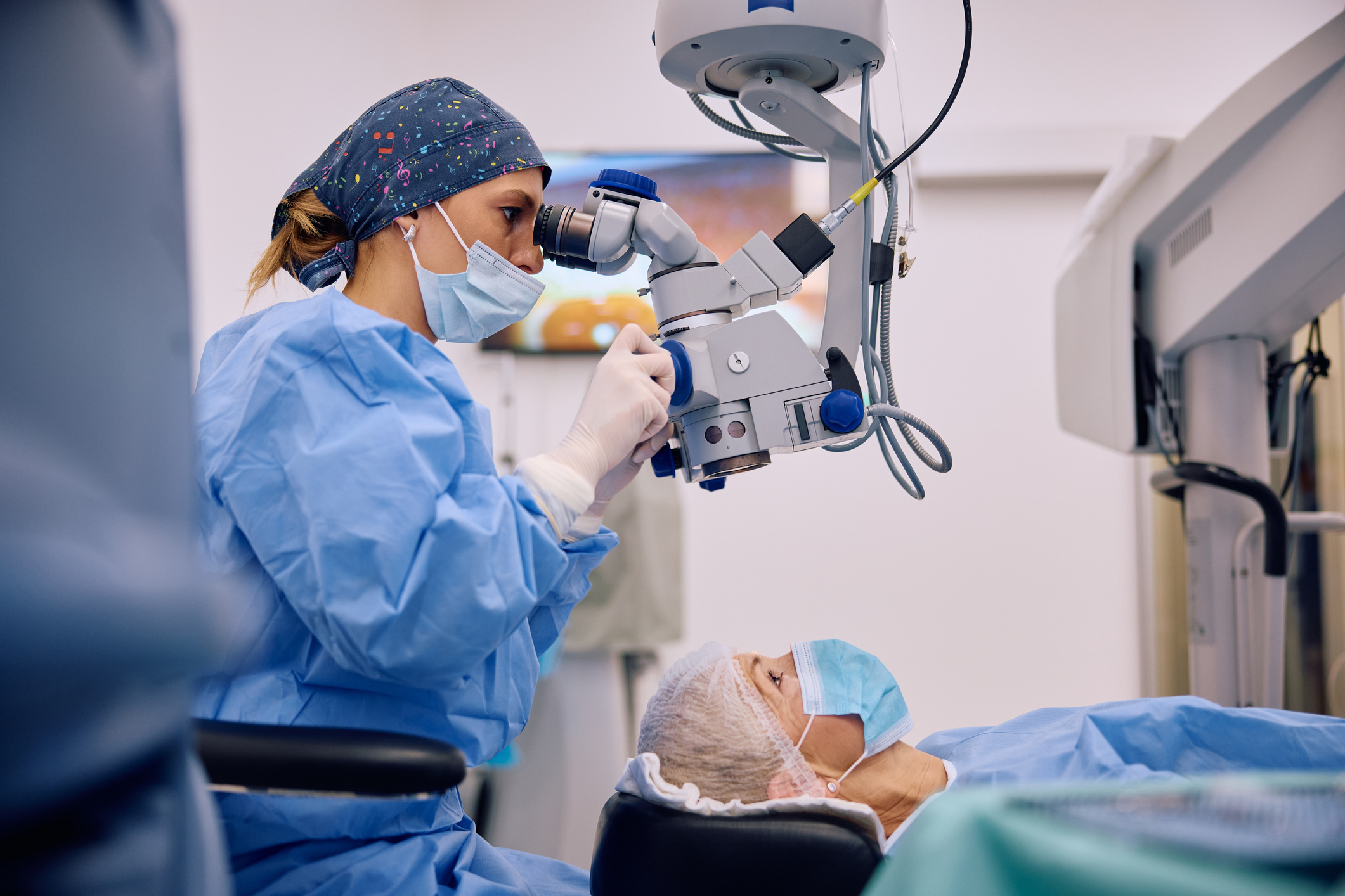
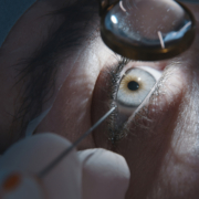
Image of an older man getting screened for glaucoma.

Cataract Surgery

If the cataract significantly impacts your vision and daily activities, cataract surgery is recommended. This involves removing the cloudy lens and replacing it with an artificial lens, also known as an intraocular lens (IOL).

Types of IOLs include:

  • Monofocal lenses – Set to focus at one distance (usually for distance vision)
  • Multifocal lenses – Allow both near and far focusing
  • Toric lenses – Correct astigmatism during surgery 
  • Light – Adjustable Lenses

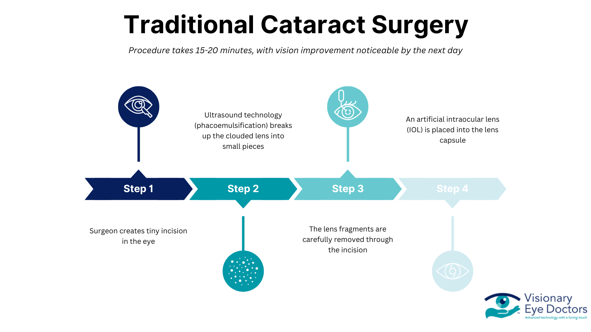
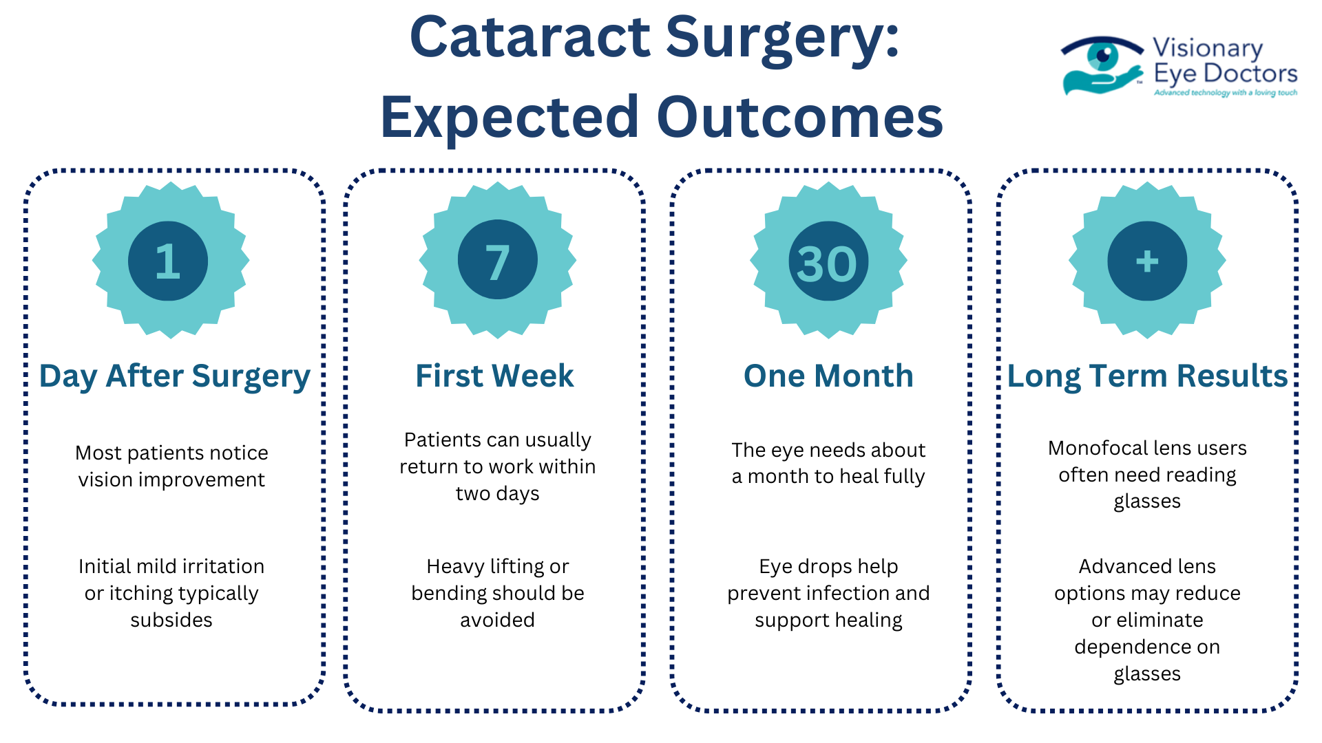
Most cataract patients report noticeable improvement in clear vision within a few days, with full recovery in 4–6 weeks.

Cataract surgery is one of the most commonly performed surgical procedures worldwide, with a success rate of over 95%. The procedure typically takes less than 30 minutes and is performed on an outpatient basis, meaning you can go home the same day. Modern techniques use ultrasound (phacoemulsification) to break up the cloudy lens before removal, requiring only tiny incisions that often heal without stitches.

Preventing Cataracts or Delaying Their Progression

While not all cataracts can be prevented, you can lower your risk by protecting your eyes and maintaining overall health:

  • Wear UV-blocking sunglasses in bright conditions
  • Stop smoking
  • Control diabetes and other chronic illnesses
  • Eat a balanced diet rich in leafy greens and antioxidants
  • Follow regular exam schedules recommended by the National Eye Institute

Certain nutrients may help protect against cataract formation. Vitamins C and E, lutein, zeaxanthin, and omega-3 fatty acids have shown promise in some studies. A diet rich in colorful fruits and vegetables provides these protective compounds naturally.

Taking Action: Restoring Your Clear Vision

Cataracts are a common part of aging, but that doesn’t mean you have to live with cloudy vision or limited night vision. Recognizing the signs of cataracts early and discussing your options with an eye doctor can help you preserve your vision and quality of life.

At Visionary Eye Doctors in Maryland and Washington, DC, we’ve helped thousands of patients regain clear vision through expert diagnosis and advanced cataract surgery using the latest intraocular lens technology.

If you’ve noticed any changes in your vision, don’t wait. Schedule your eye exam today.

Image of a mom taking her daughter to get an exam exam in the event of hereditary glaucoma.

Is Glaucoma Hereditary? Understanding Your Family Risk

The short answer: Yes, glaucoma has a strong genetic component. If you have a family member with glaucoma, your risk increases significantly..

Glaucoma affects millions of people worldwide, damaging the optic nerve through increased intraocular pressure. This pressure builds when the eye’s drainage system doesn’t allow fluid (aqueous humor) to flow out properly. What many don’t realize is that by the time noticeable symptoms appear, permanent vision loss may have already begun.

But what exactly does this mean for you and your eye health? Let’s explore the hereditary nature of glaucoma, what it means for families, and the steps you can take to protect your vision

The Genetic Risk: By the Numbers

Family history stands as one of the most significant risk factors for developing glaucoma. Primary open-angle glaucoma, the most common type, has a strong genetic component that can dramatically increase your chances of experiencing optic nerve damage and potential vision loss.

When doctors know glaucoma runs in your family, they can implement more frequent screening and earlier interventions. This is crucial since intraocular pressure can damage the optic nerve without noticeable symptoms until permanent vision loss has already begun.


Infographic about the genetic and demographic risk factors for Glaucoma.

Types of Glaucoma

Not all types of glaucoma have the same genetic component. Here’s how heredity influences different forms:

Primary Open-Angle Glaucoma (POAG)

Primary open-angle glaucoma shows the strongest genetic connection. It’s the most common type, accounting for approximately 90% of all glaucoma cases in Western countries. Research has identified several specific genes linked to POAG risk, including:

  • MYOC gene – Mutations here are found in 3-5% of POAG patients
  • OPTN gene – Associated with normal tension glaucoma
  • WDR36 gene – Linked to various forms of glaucoma

These genetic variations affect how eye pressure is regulated and how the optic nerve responds to stress. While having these genetic risk factors doesn’t guarantee you’ll develop glaucoma, it does significantly increase your likelihood.

Angle-Closure Glaucoma

Primary angle-closure glaucoma also has genetic components, particularly in certain ethnic populations. People of Asian descent have anatomical eye characteristics that can be inherited and increase the risk for this type, which can cause rapid pressure buildup and require immediate medical attention.

Congenital Glaucoma

Primary congenital glaucoma, present at birth or developing in early childhood, has the clearest inheritance pattern. This rare form is linked to mutations in the CYP1B1 gene, which affects how the eye’s drainage system develops. In many cases, both parents must carry the defective gene for a child to develop this type.


Infographic about the different genetic components of glaucoma types.

Beyond Genes: Other Risk Factors For Developing Glaucoma

While genetic predisposition creates your baseline risk, several other factors influence whether someone with a family history actually develops glaucoma:

Age

Age remains a significant risk factor for most types of glaucoma:

  • Under 40: Lower risk (except with strong genetic factors)
  • Ages 40-60: Moderate and increasing risk
  • Over 60: Highest risk, with rates four times higher than younger populations

For those with a family history, the risk curve shifts earlier, with symptoms sometimes appearing 10-15 years sooner than in the general population.

Other Medical Conditions

Several health conditions can interact with genetic factors to further increase glaucoma risk:

  • Diabetes doubles the chance of developing glaucoma and can accelerate progression
  • Hypertension affects blood flow to the optic nerve
  • Heart disease may compromise oxygen delivery to eye tissues
  • Sleep apnea has been linked to normal tension glaucoma
  • Managing these conditions becomes especially important for those with a family history of glaucoma.

Environmental Factors and Lifestyle

Even with genetic risk factors, lifestyle choices can help reduce your overall risk:

  • Regular exercise can help maintain healthy eye pressure levels
  • Smoking cessation improves blood vessel health and oxygen flow
  • Eye protection prevents injuries that could trigger secondary glaucoma
  • Limited steroid use (when medically appropriate) reduces medication-induced pressure increases
  • Proper hydration supports overall eye health


Image of an eye doctor performing an exam on a patient.

Family History: What You Need to Know and Share

Understanding your family’s eye health history can help you and your eye doctors be proactive when it comes to glaucoma development. Consider these questions:

  • Has anyone in your immediate family been diagnosed with glaucoma?
  • At what age did their glaucoma develop?
  • What type of glaucoma do they have?
  • How severe is their condition?
  • What treatments have they needed?

Create a basic family eye health tree extending to grandparents, parents, siblings, and children. Share this information with your eye doctor to help them determine your risk level and appropriate screening schedule.

Testing and Early Detection: Your Prevention Strategy

For those with a family history of glaucoma, early and regular screening becomes vital. While genetic testing can provide additional information in some cases, comprehensive eye exams remain the primary tool for detection.

When to Start Regular Screenings

If glaucoma runs in your family, follow these guidelines:

Age Group Family History Recommended Screening Frequency
Under 40 Yes Every 1-2 years
40-54 Yes Annually
55+ Yes Every 6-12 months
Any Age No Every 2-4 years (more frequently after 40)

What to Expect During a Comprehensive Eye Exam

During a comprehensive eye exam for glaucoma, your doctor will perform several key tests to assess your eye health. The process begins with tonometry to measure intraocular pressure, followed by visual field testing to check your peripheral vision for any early signs of vision loss. Your doctor will then conduct a dilated eye examination, which allows for a thorough inspection of the optic nerve to detect any signs of optic nerve damage.

For more detailed evaluation, optical coherence tomography (OCT) creates precise images of your optic nerve structure, helping identify subtle changes that might indicate glaucoma. Finally, pachymetry measures your corneal thickness, as thinner corneas can increase glaucoma risk. These non-invasive tests provide a complete picture of your eye health, making regular eye exams essential for early detection and preventing permanent vision loss.

Genetic Testing Options

For families with strong patterns of glaucoma, genetic testing offers valuable insights into your risk profile. This testing benefits those with multiple affected family members, early-onset glaucoma, or rare disease forms by identifying specific mutations and determining personal risk levels. While the process requires just a simple blood or saliva sample, it’s important to note that not all genetic factors for glaucoma are known yet. Discuss with your eye doctor whether this testing would be beneficial for your specific situation.


Infographic that outlines the criteria, benefits, and other information for genetic testing for glaucoma.

Proactive Steps to Minimize Glaucoma Risks

If your family history puts you at higher risk for glaucoma, taking proactive steps becomes essential:

Monitor Your Eye Health

  • Keep regular appointments with your eye doctor
  • Know your baseline numbers for eye pressure and visual field tests
  • Watch for warning signs between examinations

Know the Warning Signs

While early-stage glaucoma often lacks noticeable symptoms, be alert for:

  • Gradual loss of peripheral vision
  • Seeing halos around lights
  • Eye pain or severe headaches (especially with closed-angle glaucoma)
  • Redness in the eye
  • Vision changes that don’t improve

Contact your eye doctor immediately if you experience sudden vision changes or eye pain, as these could indicate acute angle-closure glaucoma requiring urgent care.

Image of a young eye doctor performing an eye exam on a patient.

Treatment Options: Early Intervention Matters

For those diagnosed with glaucoma, treatment options typically include:

  • Eye drops to lower intraocular pressure
  • Laser treatments to improve drainage
  • Minimally invasive glaucoma surgeries (MIGS)
  • Traditional surgeries like trabeculectomy for advanced cases

The key is early diagnosis, which allows for less invasive treatments and better vision preservation. This is particularly important for those with genetic risk factors, who may see more rapid progression without intervention.

Knowledge is Protection

Understanding the genetic component of glaucoma empowers you to take control of your eye health. While you can’t change your genetic makeup, knowing your family history allows you to work closely with eye care professionals to detect and manage glaucoma early,  when it matters most.

At Visionary Eye Doctors, we specialize in comprehensive glaucoma care, from advanced diagnostics to the latest treatment options. We encourage everyone, especially those with a family history of glaucoma, to schedule regular comprehensive eye exams.

Don’t wait until vision changes occur. Take this proactive step today by contacting us to schedule an appointment. Your vision is precious – let us help you protect it.

Close up of a patient receiving cataract surgery.

Does Cataract Surgery Correct Vision? ​

The Short Answer: Cataract surgery can correct vision by replacing your eye’s natural lens with an artificial intraocular lens. Modern cataract surgery offers various premium IOL options that can improve both your cataract-related vision problems and other vision issues like nearsightedness or farsightedness.

While the quick answer is yes, there’s more to the story. From how cataracts affect your sight to the types of surgeries and lenses available, understanding your options can help you make the best decision for your eyes and lifestyle. Keep reading to get a full picture of what cataract surgery involves—and how it can restore your clarity.

Understanding Cataracts and Vision Impact

What Are Cataracts?

A cataract occurs when the eye’s natural lens, which normally focuses light onto the retina for clear vision, becomes cloudy. This cloudiness happens when proteins in the lens begin to break down and change their structure, blocking light from passing properly through the eye. The result is like looking through a foggy window, making daily life activities more difficult.

People with vision problems from cataracts often notice their vision becoming increasingly blurry over time. Common signs include:

  • Frequent changes in glasses prescription
  • Seeing halos around bright lights, especially while driving at night
  • Increased glare sensitivity 
  • Difficulty reading in dim lighting
  • Colors appearing faded or yellowish

Causes and Risk Factors

Age stands as the leading cause of cataracts, with most older adults developing them as a natural part of getting older. While some people may notice changes in their 50s, cataract surgery becomes most common for those in their 70s.

According to the National Eye Institute, several other factors can increase your risk of developing cataracts:

  • Extended exposure to UV light and sunlight
  • Eye injuries or trauma
  • Use of certain medications, particularly steroids
  • Medical conditions like diabetes
  • Some people are born with cataracts, though this is rare

Surgery becomes necessary when cataracts begin to interfere with daily activities and quality of life. This typically happens when updating your glasses prescription no longer helps improve your vision, or when vision changes occur rapidly within a year. At this point, the only effective treatment is surgical removal of the clouded lens and replacement with an artificial intraocular lens.

Types of Cataract Surgery

Traditional Surgery

Technology Overview:

  • Uses phacoemulsification (ultrasound technology)

Key Steps:

  • Eye surgeon makes a tiny incision in the eye
  • Ultrasound breaks up the clouded lens into small pieces
  • Lens fragments are carefully removed through the incision
  • Artificial intraocular lens (IOL) is placed into the lens capsule

Key Benefits:

  • Remains one of the most frequently performed surgical procedures worldwide
  • Well-established procedure with extensive safety record
  • Effective removal of cataracts to improve vision
  • Compatible with various types of intraocular lenses
  • Covered by most insurance plans

Patient Considerations:

  • Outpatient procedure typically takes about 15-20 minutes
  • Patients return home the same day
  • Vision improvement is noticeable by the next day
  • Complete healing takes about a month while using prescribed eye drops

Infographic describing the steps of a traditional cataract surgery.

Laser-Assisted Surgery

Technology Overview:

  • Uses femtosecond laser (produces ultra-quick pulses in trillionths of a second)
  • Replaces several steps traditionally performed by hand

Key Benefits:

  • Creates precise incisions
  • Softens the cataract for easier removal
  • Provides accuracy for premium IOL placement (multifocal or extended depth of focus lenses)
  • Can correct astigmatism during the procedure with precise corneal incisions

Patient Considerations:

  • Not required for all patients
  • Often recommended for those choosing premium lenses
  • Beneficial for certain eye conditions
  • Recovery timeline similar to traditional surgery
  • May provide more predictable results for specific cases

Both surgical options are safe and effective. The choice between them often depends on factors like:

    • Lens choice
    • Presence of astigmatism
    • Insurance coverage. 

As with any medical procedure, your eye doctor will help determine which approach best suits your specific needs and vision goals.

Lens Options and Vision Correction

Every type of cataract surgery involves replacing your cloudy natural lens with an artificial intraocular lens (IOL). There are several IOL options available, each offering different benefits depending on your lifestyle, vision needs, and budget. Understanding these options will help you make an informed decision with your ophthalmologist about which lens is right for you.

Close up of an IOL lens for a cataract lens

Standard Monofocal Lenses

Monofocal IOLs:

Monofocal lenses represent the most common choice in cataract surgery. These artificial intraocular lenses focus light at one specific distance, typically set for clear distance vision. Patients can select whether they want their vision optimized for distance, medium-range, or close-up activities. Most people opt for distance vision correction and use reading glasses for close work.

Two Different Monofocal Lens Types:

For those interested in a different approach, monovision setup allows one eye to focus on distant objects while the other handles near vision, though this arrangement isn’t suitable for everyone.

Advanced Lens Options

Modern cataract surgery offers several sophisticated lens choices. 

Light Adjustable Lenses (Latest Technology)

The Light Adjustable Lens (LAL) is a type of intraocular lens, or IOL, that is used to replace your natural lens during cataract surgery. It is the only IOL available that is customizable after cataract surgery. These lenses are made of a photosensitive material that can be changed after cataract surgery to optimize your vision. 

Multifocal Lenses:

Multifocal lenses contain multiple focusing zones, similar to bifocal glasses, allowing clear vision at various distances. While these premium IOLs might eliminate the need for glasses completely, some patients notice halos or bright lights.

Extended Depth of Focus Lenses:

Extended depth of focus (EDOF) lenses provide a single, stretched focusing zone for better distance and intermediate vision, with fewer visual disturbances than multifocal options. For patients with astigmatism, toric lenses offer built-in correction and work with any lens platform.

Expected Outcomes

  • Most patients notice vision improvement by the day after surgery
  • The eye needs about a month to heal fully, with eye drops helping prevent infection and support healing
  • Initial mild irritation or itching typically subsides after the first night
  • Patients can usually return to work within two days
  • Heavy lifting or bending should be avoided during recovery
  • Final vision results depend on the chosen lens type:
    • Monofocal lens users often need reading glasses
    • Advanced lens options may reduce or eliminate dependence on glasses

Having realistic expectations about your selected lens type helps ensure satisfaction with the results.

Infographic about expected outcomes for cataract surgery.

Recovery and Long-term Results

Immediate Post-Surgery Period

The first 24-48 hours after cataract surgery are mild and straightforward for most cataract patients. Most people notice some itching or light irritation during the first night, but the outpatient procedure causes minimal discomfort. You’ll wear a protective shield over your eye for the first day, then only at night for a week afterward.

Your eye doctor will give you specific instructions about eye drops, which you’ll need to use for about a month to help with healing and prevent infection. During this initial period, you should avoid heavy lifting or bending down. Most people can return to daily life within one or two days, though you’ll need to stick to light activities.

Vision Stabilization

Vision improvement often begins the day after surgery. While many patients see better almost immediately, your vision may be blurry vision and fluctuate for several days as your eye heals. Full vision stabilization typically occurs within a few weeks, though this can vary among patients.

Your eye doctor will schedule follow-up appointments to monitor your healing and vision progress. The first check-up usually happens the day after surgery, with additional visits during the first month. These visits help make sure your eye is healing properly and your new lens is functioning as intended.

Long-term Benefits

Once your eye fully heals, you can expect clear vision that remains stable. Many patients report better vision than they had before their cloudy lens developed. Your artificial lens won’t develop cataracts and should last for the rest of your life.

The improvements in daily life can be remarkable. Activities are made easier, including:

  • Driving (especially at night)
  • Reading
  • Watching TV
  • Using digital devices

Many patients find they need fewer vision aids than before surgery. Depending on the type of intraocular lens chosen, some people can reduce or eliminate their dependence on glasses, though reading glasses might still be needed for close-up tasks.

Conclusion

Team of medical worker performing an eye surgery.

Modern cataract surgery is a remarkable advancement that does much more than just remove cloudy lenses. Today’s surgical options can meaningfully correct vision through precise techniques and premium IOLs. The procedure allows most cataract patients to see more clearly within days, with options ranging from monofocal lenses to advanced multifocal lenses that can reduce dependence on glasses.

When considering cataract surgery, consider:

  • Your lifestyle needs
  • Your specific vision goals
  • Solutions available for:
    • Distance vision (driving)
    • Reading vision
    • Astigmatism correction

At Visionary Eye Doctors, we understand that choosing the right cataract surgery option is an important decision. Our experienced eye surgeons combine advanced technology with personalized care to help you achieve the best possible vision outcomes. Ready to learn if cataract surgery is right for you? Contact us today to schedule a consultation and discover how we can help restore your clear vision.

Picture of a woman suffering from dry eye.

Can Dry Eyes Cause Blurry Vision?

The Short Answer: Yes, dry eyes can cause blurry vision. When your eyes lack proper lubrication from tears, the surface of your eye becomes irregular and affects how light enters your eye, resulting in unclear vision.

Your eyes rely on a consistent layer of tears to maintain sharp, clear vision. Think of tears as a smooth window that light must pass through before reaching the sensitive parts of your eye. When that window becomes disrupted due to dry eyes, your vision can become temporarily blurred or distorted.

According to the National Eye Institute, dry eye affects millions of Americans, with nearly 16 million adults diagnosed with the condition. This common problem can significantly impact daily vision and comfort.

In this article, you’ll discover how tears affect your vision, what causes dry eyes to blur your sight, and practical ways to address this common problem. We’ll explore both quick fixes and long-term solutions to help you maintain clear, comfortable vision throughout your day.

What Is Dry Eye?

Dry eye syndrome often develops in three main ways:

  1. Your eyes might not produce enough tears, a common condition as people age or take certain medications. 
  2. Tears might evaporate too quickly, especially if the meibomian gland function is impaired. 
  3. Your tears may be missing important components, meaning they are unable to properly coat your eye surface. 

When any of these problems occur, your tear film becomes unstable and breaks up too quickly between blinks, leaving dry spots that can cause both vision problems and discomfort.

Why Does Dry Eye Affect Vision?

Normal Tear Function

Tears do much more than show emotion – they’re necessary for clear vision and comfort. Every time you blink, tears spread across your cornea (the clear front surface of your eye) creating a smooth surface that allows light to focus properly. 

This tear film also 

  • Washes away dust and debris
  • Prevents infection
  • Supplies oxygen and nutrients to your cornea. 

Without this constant moisturizing and protection, dry eye symptoms can cause blurry vision.

Image of a green eye producing tears.

Signs that Dry Eye is Affecting Your Vision

Common Vision Symptoms

Vision Effects

  • Dry eye syndrome directly impacts visual clarity
  • Insufficient tears lead to noticeable blurry vision throughout the day
  • Tear film functions like a camera lens:
    • Needs to be smooth and clear for sharp vision
  • Vision often fluctuates between clear and blurred as tear film breaks down and reforms

Associated Symptoms

  • Light sensitivity becomes common:
    • Bright indoor lighting causes discomfort
    • Sunlight may feel uncomfortable
  • Eye strain develops more quickly:
    • Digital device use becomes more difficult
    • Computer screen viewing is more challenging
    • Occurs because protective tear production isn’t functioning properly

Infographic about how dry eyes effect vision.

The Physical Connection

The Root Cause: Corneal Surface Changes

  • The cornea (clear front surface of your eye) requires constant tear production
  • Healthy tears keep the cornea smooth and transparent
  • When tears are lacking, the corneal surface becomes irregular and rough
  • This irregular surface scatters incoming light instead of focusing it properly

These corneal surface irregularities directly correlate with decreased visual performance in dry eye patients.

Why Vision Problems Fluctuate

  • Blinking temporarily spreads fresh tears across the cornea
  • This momentarily improves visual clarity
  • Vision quickly becomes distorted again when:
    • Tears evaporate too quickly
    • Tear composition isn’t the right consistency

This explains why dry eye-related vision problems tend to come and go throughout the day.

Warning Signs to Watch For

Pay attention if you notice:

  • Vision that changes between clear and blurry throughout the day
  • Difficulty focusing when using digital devices
  • Eyes that feel gritty or scratchy
  • Burning or stinging sensations
  • Watery eyes or excessive tearing
  • Redness in the whites of your eyes

See an eye doctor promptly if these dry eye symptoms persist for more than a few days, especially if artificial tears don’t provide relief. Early treatment can prevent corneal damage and long-term vision problems. Your eye doctor can determine the underlying cause and recommend the most effective treatment options for your specific situation.

Causes and Risk Factors

Image of two woman with faces next to each other showing their eyes.

Common Causes

As we age, our eyes naturally produce fewer tears, with most people over 65 experiencing dry eye symptoms. 

According to the American Academy of Ophthalmology, several health conditions can trigger dry eye syndrome, including:

  • Rheumatoid arthritis
  • Diabetes
  • Autoimmune disease. 

The AAO also notes that taking these medications may reduce tear production:

  • Antihistamines
  • Decongestants
  • Blood pressure medications
  • Antidepressants 

Hormonal changes in women, particularly during pregnancy and menopause, can also lead to chronic dry eye.

Environmental Factors

  • Extended computer screen time can significantly impact tear film, as people tend to blink less frequently when looking at digital devices
  • Dry or windy climates speed up tear evaporation
  • Indoor environments with air conditioning or heating can decrease air humidity 
  • Smoke exposure and high altitudes 
  • Poor air quality in office spaces or other workplace settings

Lifestyle Contributors

  • Wearing contact lenses for long periods can interfere with the natural tear film across the eye surface
  • Not drinking enough water throughout the day leads to dehydration, which affects tear production
  • Poor sleep habits can impact overall eye health and vision
  • Activities requiring intense focus often result in reduced blinking, which disrupts the eye’s natural moisturizing process 

Making lifestyle changes to daily routines, like taking regular screen breaks and staying hydrated, can help maintain better eye moisture levels.

Conclusion and Solutions

Dry eye syndrome can definitely cause blurry vision, but there are many effective treatment options to address this common condition. Proper tear production and tear film quality are fundamental for clear, comfortable vision. When these are disrupted, both your eye health and visual clarity can suffer.

Treatment Options

  1. Over-the-counter artificial tears provide quick relief for mild dry eye symptoms. Look for preservative-free eye drops to minimize irritation. 
  2. Lifestyle changes like using a humidifier, taking regular computer screen breaks, and staying hydrated can help maintain tear film. 

3. For more significant cases, prescription medications, tear duct plugs, or specialized treatments may be needed to increase tear production and reduce inflammation.

Infographic about dry eye treatment options.

When to Schedule an Eye Exam

Schedule an appointment with Visionary Eye Doctors if you experience:

  • Persistent blurry vision that doesn’t clear with blinking
  • Dry eye symptoms that interfere with daily life
  • Symptoms that don’t improve with over-the-counter drops

Our experienced eye doctors at Visionary Eye Doctors can evaluate your tear production and quality to create a personalized treatment plan. Contact us today to take the first step toward relief from chronic dry eye and blurred vision. Restore your eye comfort and clear vision!