Toddler child pointing to a vision chart during eye check

Back-to-School Myopia Management for Kids: What Parents Should Know

At a Glance: Myopia management uses proven treatments like special soft contact lenses, ortho-k lenses, and low-dose atropine drops to slow how quickly a child’s nearsightedness gets worse. Starting early, around the back-to-school season, gives your child the best shot at clear vision now and a lower risk of serious eye problems as an adult.

The new school year often brings new vision changes. Kids spend more hours reading, writing, and looking at screens, and parents start to notice squinting or complaints about the board at school. If your child is nearsighted, the goal is to correct their vision today and to slow myopia progression over time. 

What Is Myopia, and Why Are More Kids Getting It?

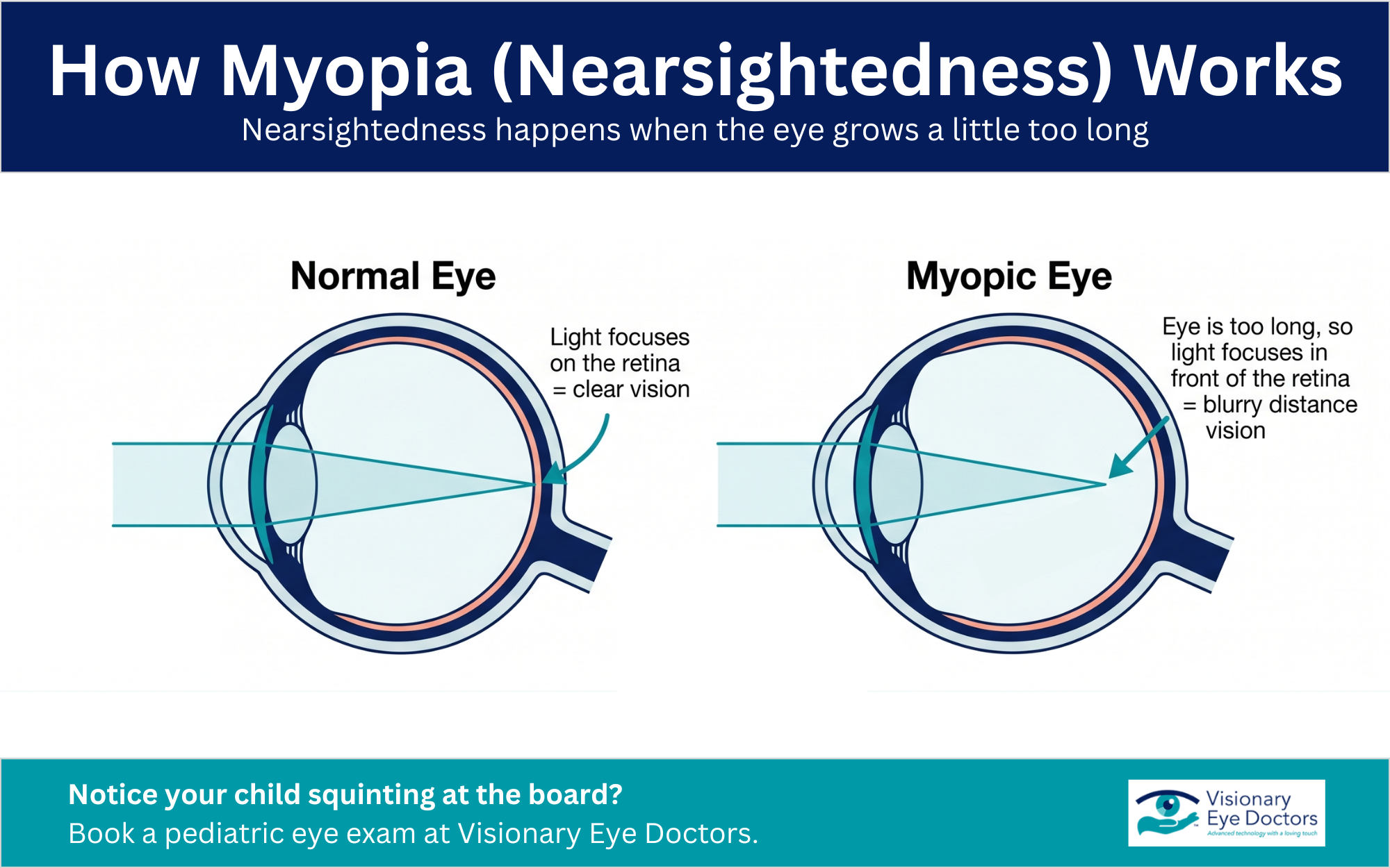
Myopia, also called nearsightedness, is a common refractive error. The eye grows a little too long, so light focuses in front of the retina instead of on it. That leads to blurry distance vision while close-up objects stay clear.

Infographic: How Myopia (Nearsightedness) Works

Childhood myopia is rising fast. Researchers project that nearly half of the world’s population could be nearsighted by 2050, and rates among children keep climbing.

The main risk factors for childhood myopia include:

  • Genetics. A child with one or two nearsighted parents has a higher risk.
  • Lifestyle. More time on near work and digital devices, plus less time outdoors, is linked to more myopia in younger children.

Why the Back-to-School Season Matters

Myopia tends to start and speed up during the school years. A child who becomes nearsighted early often ends up with a stronger prescription as they grow.

Back-to-school is a smart time for a regular eye exam because:

  • Vision may have changed over summer break.
  • Catching myopia early opens up more treatment options.
  • A clear baseline lets your eye doctor track myopia progression at follow-up visits.

Signs Your Child May Be Nearsighted

Kids do not always say, “I can’t see.” Watch for these clues:

  • Sitting very close to the TV or holding devices near the face
  • Squinting to make out distant objects or the whiteboard
  • Frequent headaches or eye rubbing
  • Trouble in sports that require clear distance vision
  • Slipping grades or losing interest in classroom work

If you notice any of these, schedule pediatric eye care. Active kids especially benefit from clear vision on the field and in the classroom.

The Risks of Letting Myopia Progress

Glasses correct vision, but needing stronger glasses every year can signal that the eye keeps growing. When progressive myopia continues into the teen years, it can reach high myopia. High myopia raises the lifetime risk of serious eye conditions, including:

  • Retinal detachment
  • Macular degeneration (the myopic form)
  • Glaucoma and early cataracts

That is why slowing myopia matters. Myopia control helps protect your child’s long-term eye health while it sharpens their vision today. (Learn more about the early signs of glaucoma.)

Myopia Treatment Options That Slow Progression

Several evidence-based treatments can slow progression. Your eye doctor will match the option to your child’s age, prescription, and daily routine.

Treatment How It Works Good to Know
Stellest lenses FDA-authorized eyeglass lenses for kids 6 to 12 that look like regular glasses. A clear center keeps distance vision sharp, while tiny rings around the edge gently prompt the eye to slow its growth A good fit for younger children and kids not ready for contacts; a two-year study showed about 71% slower myopia progression versus standard glasses
MiSight lenses The first FDA-approved soft contact lenses for kids 8 to 12, these daily soft contact lenses correct blurry distance vision and slow eye growth Single-use, so no cleaning is needed; research finds dual-focus soft lenses slow progression by roughly 50% to 60% over two to three years
Ortho-k lenses Rigid lenses worn overnight gently reshape the cornea while your child sleeps Your child goes glasses-free and contact-free during the day
Low-dose atropine A nightly eye drop that helps slow myopia progression Often most helpful in younger children; used with close monitoring
Multifocal soft contact lenses A soft contact lens with a clear center for distance and outer rings that slow eye growth A good match for many myopic children

Each plan includes ongoing monitoring so your eye doctor can adjust as your child grows. No single option is right for every child, which is why a personalized fit matters.

Simple Habits That Support Your Child’s Vision

Daily habits will not replace treatment, but they help:

  • Get outside. Time in natural daylight is linked to slower myopia in kids. Aim for outdoor play most days.
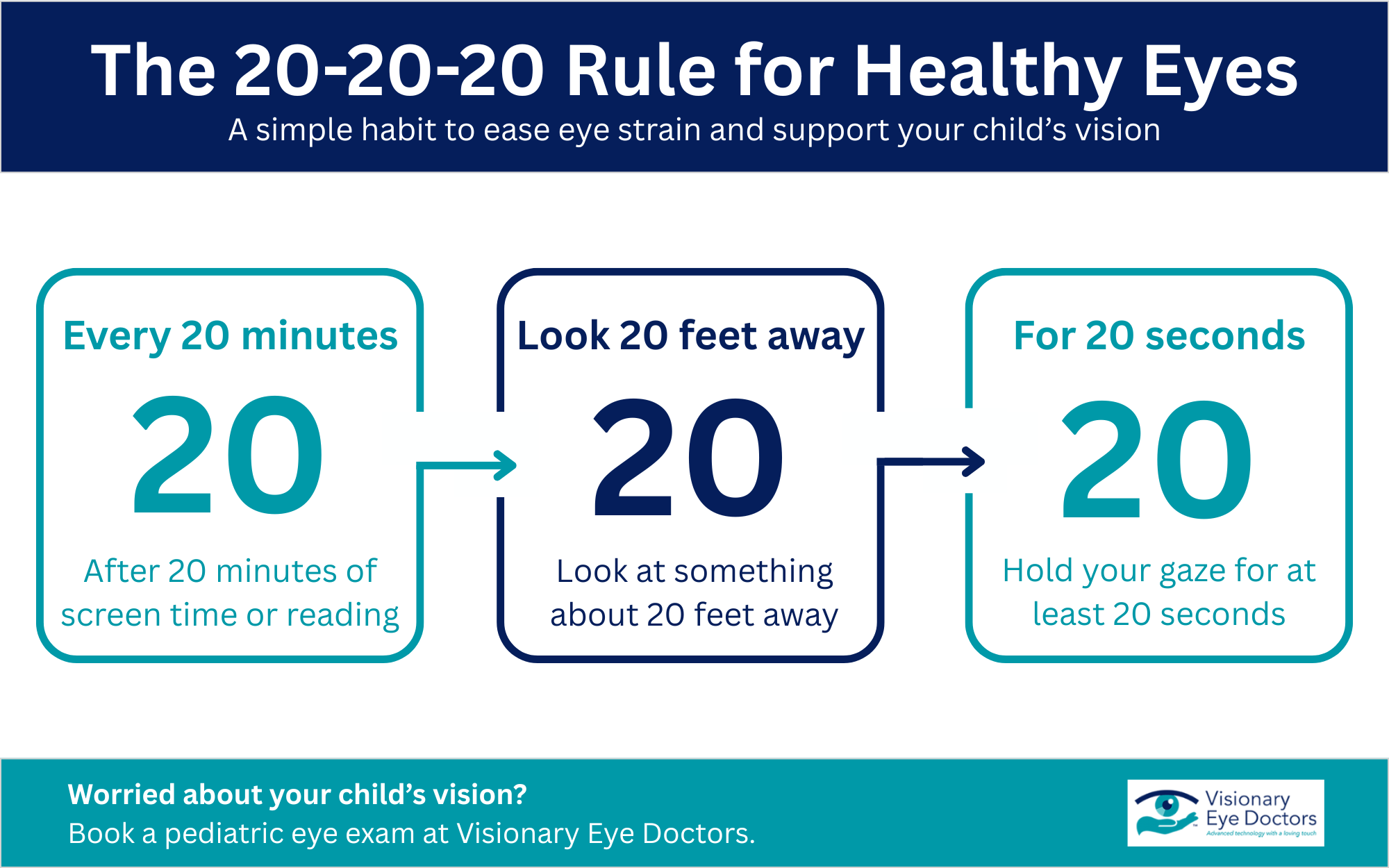
  • Take regular breaks. Try the 20-20-20 rule: every 20 minutes, look at something about 20 feet away for 20 seconds.
  • Manage screen time. Set limits on digital devices and keep screens at a comfortable distance.
  • Use good lighting. Bright, even light takes strain off your child’s eyes during reading and homework.

Infographic: The 20-20-20 Rule for Healthy Eyes

What to Expect at a Myopia Management Visit

A myopia-focused exam goes beyond a standard vision check. Here is the typical path:

  • Full eye exam. We measure the refractive error and check overall eye health.
  • Myopia tracking. We may measure the length of the eye to see how fast it is changing.
  • A personalized plan. We walk through treatment options and pick what fits your child’s needs.
  • Follow-up visits. Short, regular checks show us what is working so we can fine-tune the plan.

Common Questions Parents Ask

At what age should myopia management start?

Sooner is better. Myopia control often works best in younger children, since they have the most eye growth still ahead of them. If your child’s prescription climbs each year, ask your eye doctor whether a plan makes sense now rather than waiting.

Will my child still need glasses?

Most likely, yes. These treatments slow myopia progression, but they do not reverse it. The aim is to keep the prescription from rising as fast, which lowers the chance of high myopia and the eye problems tied to it later in life.

Does myopia ever stop getting worse?

For most kids, myopia slows down and settles in the late teen years or early adulthood. Treatment during the school years helps limit how far it goes, which protects vision well into the young adult years.

Give Your Child a Clear Start to the School Year

Young girl looking closely at book while wearing glasses

Myopia in children rarely stands still, but you can slow it down. With an early eye exam and a plan built around your child, you protect their clear vision today and lower the risk of serious eye problems later in life. The back-to-school season is a natural time to begin.

At Visionary Eye Doctors, we offer myopia management for families across Washington, DC, and Maryland. Our team fits soft contact lenses, ortho-k lenses, and other myopia control options, then supports your child with caring, ongoing monitoring at every follow-up. We explain each step in plain language so you always know what comes next.

Give your child the gift of clear vision this school year. Book an appointment today and let us build a myopia management plan that grows with them.

girlvisiting oculist for measuring eyesight with phoropter

When Should Kids Get an Eye Exam?

The Short Answer: Children should have their first eye exam around 6 months of age, another between ages 3 and 5, and then annual eye exams once they start school. Early detection of vision problems supports learning, development, and long-term eye health.

Parents often wonder if their child needs a pediatric eye exam, especially when the school nurse already runs basic vision screenings. The truth is that screenings catch only a fraction of vision issues. A pediatric eye exam looks much deeper at your child’s eyesight, eye coordination, and overall eye health. Catching a refractive error or lazy eye early can change a child’s whole school experience.

Recommended Eye Exam Schedule by Age

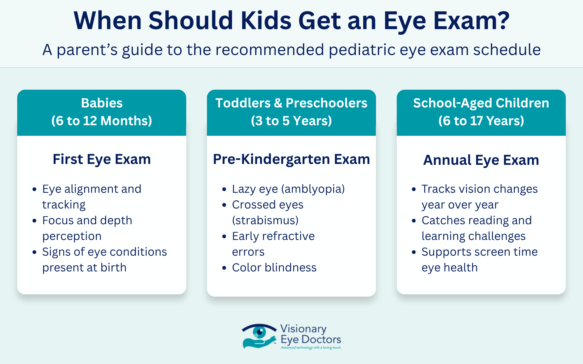
The American Optometric Association suggests a specific timeline for children’s eyes. Here is what regular pediatric eye exams look like at each stage.

Infographic: When Should Kids Get an Eye Exam?

Babies (6 to 12 Months)

A baby’s eyes develop quickly during the first year. A first eye exam at this age checks:

  • Eye alignment and tracking
  • Focus and depth perception
  • Signs of any eye conditions present at birth

Toddlers and Preschoolers (3 to 5 Years)

Between the ages 3 and 5, kids should have another exam before kindergarten. This visit looks for:

  • Lazy eye (amblyopia)
  • Crossed eyes (strabismus)
  • Early refractive error, like nearsightedness or farsightedness
  • Color blindness

School-Aged Children (6 to 17 Years)

Once a child starts school, an annual eye exam is the standard recommendation. Reading, writing, and screen use all place new demands on a child’s vision, so regular exams help track changes year over year.

Why a School Vision Screening Is Not Enough

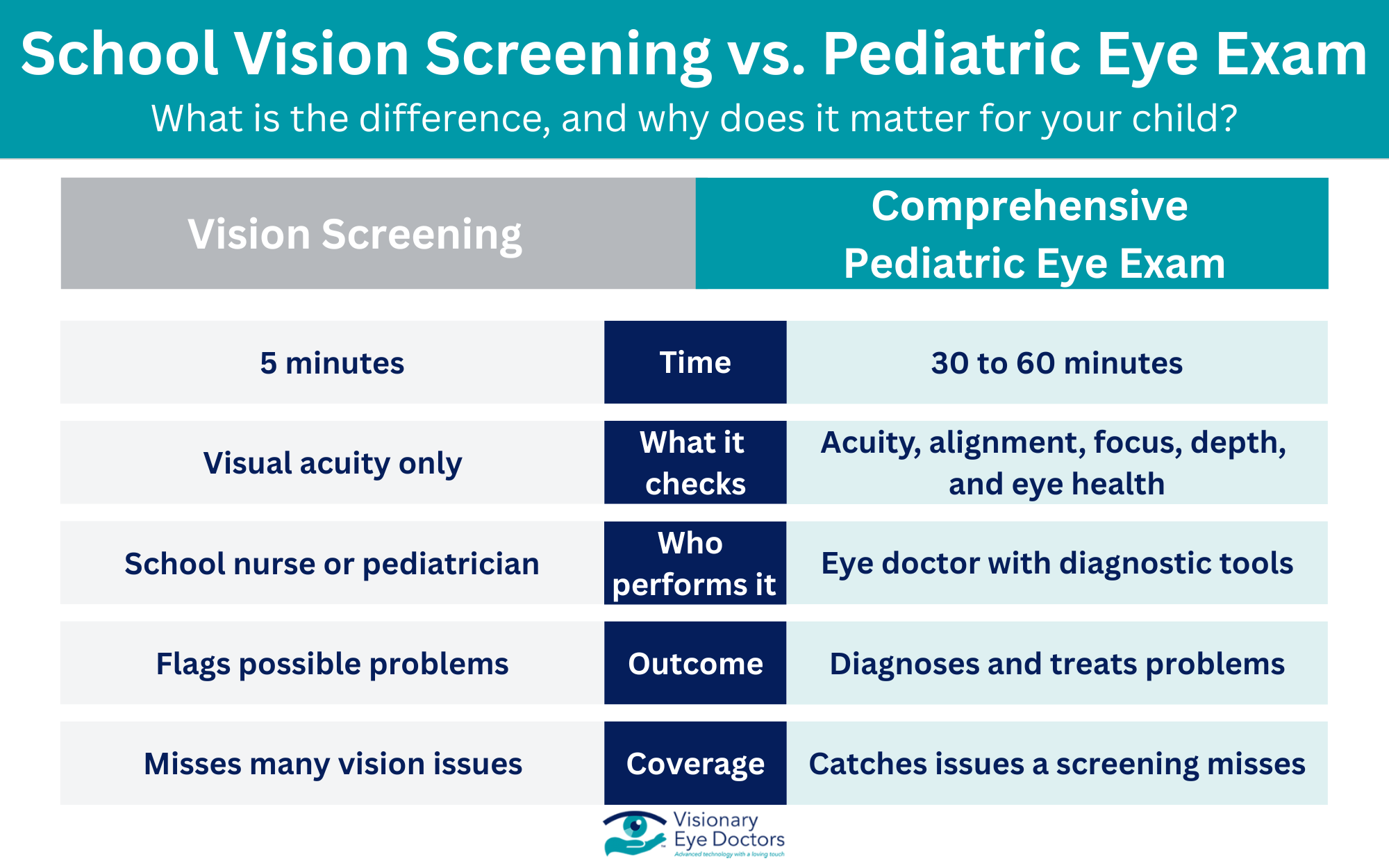
Many parents assume the quick chart test at school covers everything. Basic vision screenings mostly measure visual acuity, which means how clearly a child can see letters on a chart from across the room. They miss a lot.

A full pediatric eye exam also checks:

  • Binocular vision (how well the eyes work together)
  • Depth perception
  • Eye focusing and tracking
  • Eye health inside and out
  • Visual skills tied to reading and learning

Infographic: School Vision Screening vs. Pediatric Eye Exam

The American Academy of Ophthalmology notes that a child can pass a screening and still have a real vision problem affecting school performance.

Signs Your Child May Have a Vision Issue

Younger kids often do not realize their eyesight is not normal. They assume everyone sees the way they do. Watch for these signs:
  • Squinting or tilting the head to see
  • Sitting very close to the TV or holding devices near the face
  • Frequent eye rubbing
  • Trouble reading or skipping lines while reading
  • Short attention span during close-up tasks
  • Headaches after schoolwork
  • Avoiding reading, puzzles, or other detail-heavy activities
  • One eye that drifts or turns inward or outward

Many obvious symptoms show up at home before they show up in the classroom. If you notice any of these, schedule an appointment today rather than waiting for the next checkup.

Common Eye Problems in Children

A kid’s eye exam can pick up several conditions. Some of the most common include:

Condition What It Is
Refractive error Nearsightedness, farsightedness, or astigmatism
Amblyopia (lazy eye) One eye does not develop normal vision; affects 2 to 3 percent of children
Strabismus The eyes do not line up together
Convergence insufficiency The eyes struggle to focus together on near objects
Color blindness Trouble telling certain colors apart

Early detection matters most for lazy eye. The visual abilities of children develop fastest before age 7, so treatment works best when started young. Vision therapy, corrective lenses, or patching can correct many of these eye problems when caught early.

What Happens at a Pediatric Eye Exam

Parents sometimes worry their child is too young for a real exam. A pediatric optometrist or pediatric ophthalmologist uses age-appropriate tools, including pictures and lights, for kids who cannot read yet.

A typical visit includes:

  • A review of family eye history and any concerns
  • Tests for visual acuity using letters, shapes, or pictures
  • Checks for eye alignment, focusing, and depth perception
  • A look at eye health using gentle lights and lenses
  • A discussion of glasses, contact lenses, or vision therapy if needed

The whole exam usually takes 30 to 60 minutes. 

Screen Time and Your Child’s Eyes

Screen time has changed the way children’s eyes work day to day. Long hours on phones, tablets, and laptops can lead to:

  • Eye strain and tired eyes
  • Dry eyes from less blinking
  • Headaches
  • Faster progression of nearsightedness in some kids

A simple habit called the 20-20-20 rule helps. Every 20 minutes, look at something 20 feet away for 20 seconds. Outdoor play also supports healthy vision in young children. Even with good habits, regular exams remain the best way to track changes in a child’s eye health.

When Do Kids Need Glasses or Contact Lenses?

If an exam shows a refractive error, your eye doctor will talk through vision correction options. Glasses are usually the first step for younger children, and frames have come a long way in terms of durability and style. Older children and teens who are responsible enough to handle daily care may move into contact lenses. The right choice depends on lifestyle, prescription, sports involvement, and how well a child can stick to a cleaning routine.

For kids with stronger prescriptions or fast-changing eyesight, your eye doctor may also discuss myopia management options that can slow the progression of nearsightedness over time.

Supporting Your Child’s Vision Starts Here

Regular eye exams give kids their best shot at clear vision, strong school performance, and lifelong eye health. Starting at 6 months, then again before kindergarten, and once a year through the school years, these visits catch problems that basic vision screenings miss. From lazy eye to early refractive errors, early detection often makes treatment simpler and more effective.

Visionary Eye Doctors provides pediatric eye exams for families throughout Washington, DC and Maryland. Our team examines your child’s eye health, eye coordination, and visual skills using advanced diagnostics, and we take the time to explain what we find in language that makes sense.

Help your child start the school year with clear vision. Book an appointment today with our team.

woman itching eyes at laptop

What Causes Dry Eyes All of a Sudden?

The Short Answer: Sudden dry eye usually happens when something disrupts your tear film, whether that’s dry air, screen time, a new medication, or an underlying health issue. Most cases clear up with a few simple home changes, though lingering symptoms call for a visit to your eye doctor.

One morning, your eyes feel fine. By afternoon, they’re gritty, red, and stinging every time you blink. Sudden dry eye can feel confusing, especially if you’ve never dealt with it before. The good news is that most causes are treatable once you know what’s behind them.

How Your Eyes Stay Moist

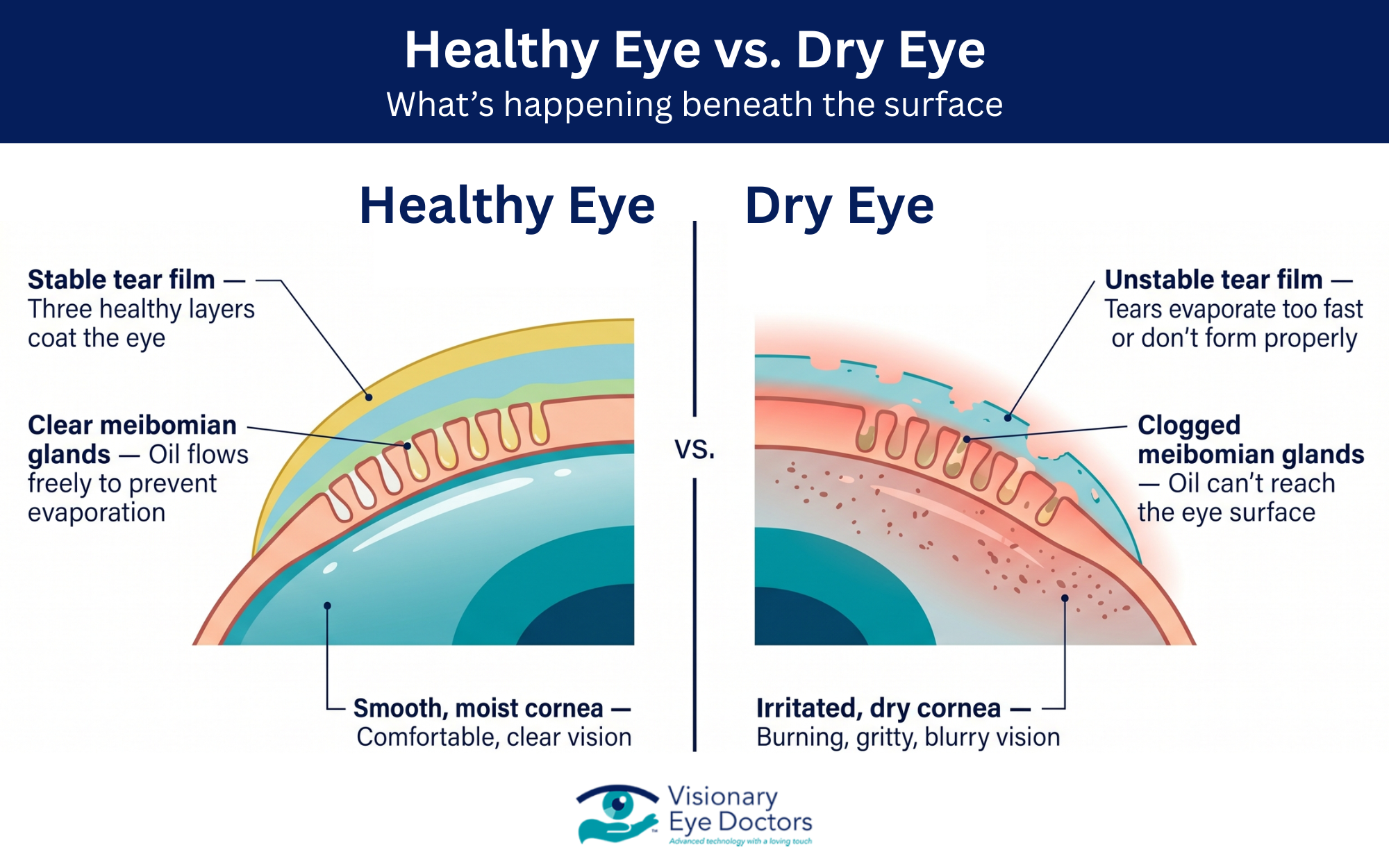
Before looking at causes, it helps to know how tears work. Your eyes rely on three layers that make up the tear film:

  • Oily layer: Produced by the meibomian glands in your eyelids, this slows tear evaporation.
  • Watery layer: Made by the lacrimal gland, this keeps your eye surface hydrated.
  • Mucus layer: Helps tears spread evenly across your eye.

Tears also drain through tiny tear ducts in the inner corners of your eyes. When any part of this system falls out of balance, dry eye symptoms show up fast.

Common Causes of Sudden Dry Eye

Dry eyes don’t always have one single trigger. Often, several external factors and daily habits stack up at once.

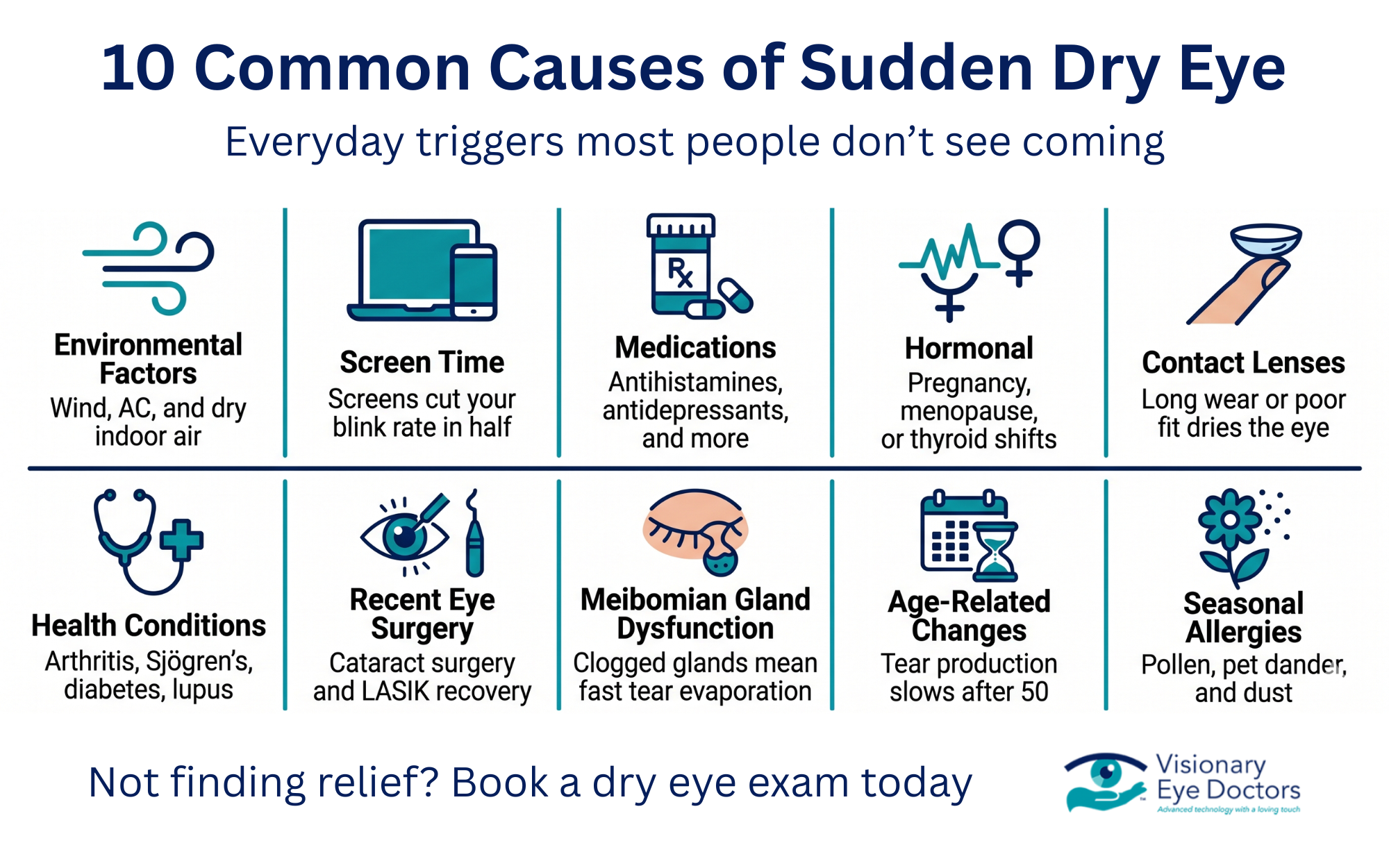
infographic: 10 common causes of sudden dry eye

1. Environmental Factors

Weather and indoor air can dry out your eyes in hours. Common culprits include:

  • Windy or low-humidity days
  • Air conditioning or forced-air heating
  • Smoke from fires or candles
  • High altitude or airline cabins
  • Ceiling fans blowing directly on your face

2. Screen Time and Daily Habits

Staring at a phone or monitor slows your blink rate by about half. Fewer blinks mean faster tear evaporation. If your daily routine involves long stretches at a computer, sudden dry eye may catch up with you during a busy week.

3. Seasonal Allergies and Irritants

Seasonal allergies, pet dander, dust, and pollen can all trigger dryness. Allergic conjunctivitis often feels similar to dry eye disease, with itching, redness, and watery eyes showing up together. Oral antihistamines help allergy symptoms but can actually reduce tear production as a side effect.

4. Prescription Medications

Many prescription medications list dry eye as a possible side effect. Ask your doctor if you recently started:

  • Antihistamines (oral or nasal)
  • Antidepressants
  • Blood pressure medications
  • Birth control pills
  • Acne treatments like isotretinoin

5. Hormonal Changes

Hormonal changes during pregnancy, menopause, or thyroid shifts can reduce tear production. Women are more likely to experience sudden dry eye during these life stages.

6. Contact Lenses

Contact lenses absorb moisture from your eye and can worsen dryness, especially if worn too long or if the fit isn’t right. Switching brands, sleeping in lenses, or skipping replacements can trigger symptoms overnight.

7. Health Conditions

Sometimes dry eye points to a larger issue. Health conditions linked to dry eye include:

If you have one of these conditions and notice new eye symptoms, mention it to your eye doctor during your next visit.

8. Recent Eye Surgery

Cataract surgery and LASIK can both cause temporary dryness while your eyes heal. Symptoms usually fade over weeks to months as the surface of the eye settles.

9. Meibomian Gland Dysfunction

When the meibomian glands get clogged, the oily layer thins out and tears evaporate too fast. Meibomian gland dysfunction is one of the most common potential causes of both chronic and sudden dry eye syndrome.

10. Age-Related Changes

Tear production naturally slows with age, especially after 50. If dry eye feels new in your 50s or 60s, small lifestyle changes may help a lot, but a full exam can also rule out age-related conditions like meibomian gland dysfunction or other eye health issues that become more common over time.

Symptoms to Watch For

Dry eye shows up in more ways than you might expect. Common dry eye symptoms include:

  • Burning, stinging, or gritty feeling
  • Redness or irritation
  • Blurry vision that clears with a blink
  • Sensitivity to light
  • Stringy mucus in or around the eyes
  • Watery eyes
  • Trouble wearing contact lenses

Watery eyes may sound like the opposite of dry eye, but when your eye surface gets irritated, your body releases reflex tears that don’t have the right makeup to actually soothe it.

When Dry Eye Feels Like Something Else

Blurred vision, floaters, or flashes of light can mimic dry eye but may point to other conditions that affect the optic nerve or retina, such as retinal detachment. If vision changes feel sudden or severe, call your eye doctor right away rather than waiting to see if they pass.

infographic: healthy eye vs dry eye

Home Remedies and Simple Changes

Many cases of sudden dry eye respond well to home remedies and lifestyle changes. Try these first for better eye comfort.

Quick Fixes

  • Use preservative-free artificial tears or over-the-counter eye drops a few times per day
  • Apply a warm compress to closed eyes for 5 to 10 minutes to loosen oil in the meibomian gland openings
  • Run a humidifier in rooms where you spend the most time
  • Wear wraparound sunglasses outdoors to block wind and sun
  • Follow the 20-20-20 rule: every 20 minutes, look 20 feet away for 20 seconds

Diet and Hydration

  • Drink water steadily throughout the day
  • Add omega-3 fatty acids from fish, flaxseed, or walnuts
  • Limit alcohol and caffeine, both of which can dehydrate you

Contact Lens Care

  • Take breaks with glasses, especially on long workdays
  • Replace lenses on the schedule your doctor recommends
  • Ask about lens types designed for dry eye

When to See an Eye Doctor

If symptoms last more than a week or two, or if they get worse during daily activities, it’s time for a comprehensive eye exam. Signs that warrant a visit include:

  • Pain or redness that won’t go away
  • Vision that stays blurry after blinking
  • Discharge or crusting
  • Trouble wearing contact lenses
  • Symptoms that start after beginning a new medication

At Visionary Eye Doctors, we look at every layer of the tear film, check your meibomian glands, and rule out other eye health concerns. Depending on what we find, treatment may include prescription eye drops, in-office gland expression, punctal plugs, or small adjustments to your daily habits. Our Corneal Care Center handles everything from mild irritation to advanced dry eye disease.

What to Expect at Your Appointment

A dry eye visit goes beyond a standard vision check. Here’s what usually happens:

  • A review of your symptoms, medications, and daily routine
  • Measurement of tear volume and tear evaporation rates
  • A close look at the meibomian gland openings and oil flow
  • A check for surface damage, inflammation, or signs of autoimmune disease
  • A discussion of treatment options matched to your results

Most exams take under an hour, and many patients leave with a plan they can start the same day.

Find Relief with Visionary Eye Doctors

woman putting in eye drops to relieve dry eye

Sudden dry eye can throw off your whole day, from reading to driving to wearing contacts. The good news is that most cases improve once the cause is known. Small steps like adding omega-3 fatty acids, using artificial tears, and limiting screen time often make a real difference within days.

If those simple changes aren’t producing enough tears to keep you comfortable, our team at Visionary Eye Doctors in Washington, DC, and Maryland is here to help. We’ll look at the full picture, from contact lens habits to any medications or health conditions that may be involved, and build a plan that fits your lifestyle. Book an appointment today and get back to a comfortable vision.

Allergies and Itchy Eyes: Causes, Triggers, and What Actually Helps

The Short Answer: Eye allergies happen when your immune system overreacts to airborne allergens like pollen or pet dander, triggering symptoms like itchy, watery, or red eyes. Most cases respond well to the right treatment, but knowing what’s causing your symptoms is the first step to real relief.

If your eyes are constantly itchy, watery, or irritated during allergy season, you’re not alone. Eye allergy symptoms affect millions of people every year, and they can make daily life genuinely miserable. The tricky part is that not all red, irritated eyes are the same. Understanding what’s actually going on helps you treat the right thing, not just the symptom.

What Is an Eye Allergy?

An eye allergy, also called allergic conjunctivitis, happens when your immune system reacts to something that isn’t actually a threat. When an allergen lands on the surface of your eye, your body releases histamine from mast cells. That chemical response is what causes swelling, redness, and that relentless itchy eye feeling.

There are a few different types:

  • Seasonal allergic conjunctivitis is the most common. It flares during pollen season, usually in the spring and fall.
  • Perennial allergic conjunctivitis happens year-round, typically triggered by indoor allergens.
  • Atopic keratoconjunctivitis is a more serious, chronic form that can affect your cornea if left untreated.

Common Allergens and Triggers

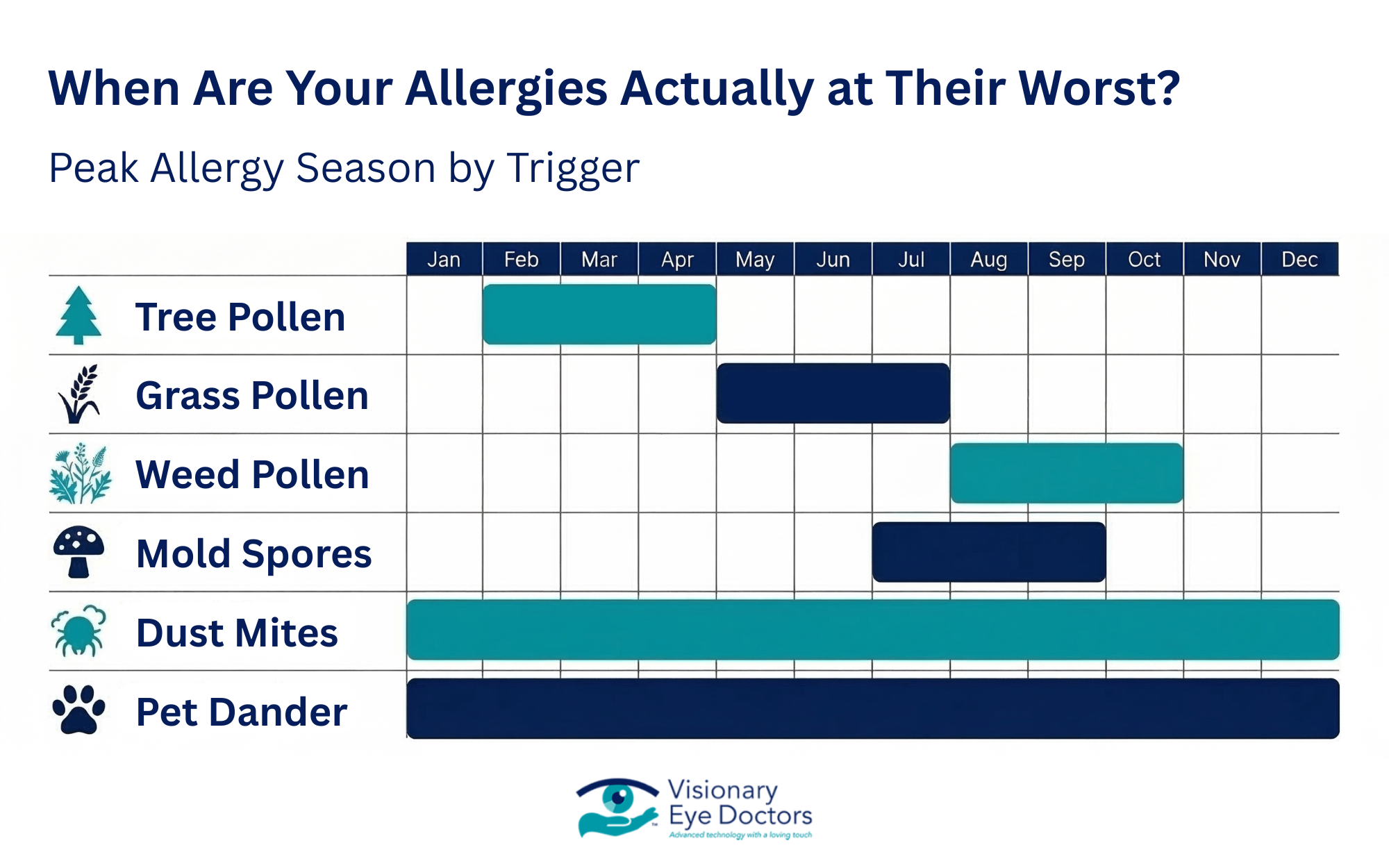
Infographic: when are your allergies actually at their worst?

Eye allergy symptoms can be triggered by both outdoor and indoor sources. The most common allergen categories include:

Outdoor triggers:

  • Tree, grass, and weed pollen (the main driver of hay fever and seasonal allergy flare-ups)
  • Mold spores in damp outdoor environments

Indoor triggers:

  • Pet dander from cats, dogs, and other animals
  • Dust mites in bedding, carpets, and upholstered furniture
  • Mold growing in humid areas of your home

During high pollen count days, symptoms tend to spike. The Asthma and Allergy Foundation of America tracks seasonal allergy data and pollen forecasts that can help you plan outdoor time during peak pollen season.

Eye Allergy vs. Pink Eye vs. Dry Eye

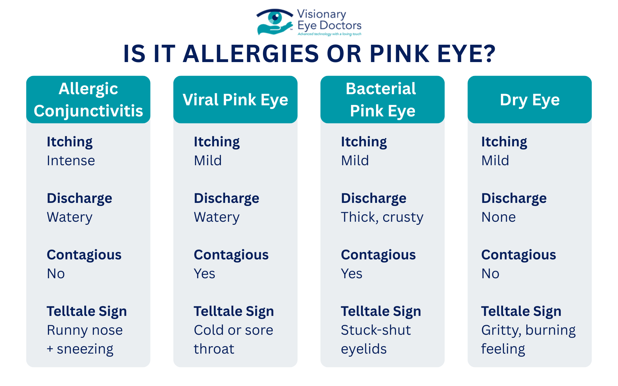
One of the most common points of confusion is telling eye allergy symptoms apart from other conditions with similar symptoms.

Allergic Conjunctivitis

  • Itchy eyes, watery eyes, redness, watery discharge
  • Not contagious
  • Almost always paired with an itchy nose, runny nose, or sneezing

Viral Conjunctivitis (Pink Eye)

  • Red eye, watery, burning sensation
  • May come with a sore throat or cold-like symptoms
  • Contagious

Bacterial Infections (Pink Eye)

  • Thick or yellow-green discharge, crusty eyelids, red eye
  • Contagious
  • Requires antibiotic treatment

Dry Eye Syndrome

  • Burning, gritty feeling, fluctuating vision
  • Not contagious
  • Can overlap with and worsen allergy symptoms

Infographic: is it allergies or pink eye?

That last point matters. Some people deal with both dry eye and eye allergies at the same time, and the symptoms overlap in ways that make it easy to misread what’s happening. An eye doctor can help sort out what’s actually going on.

What Actually Helps For Allergies

Antihistamine Eye Drops

Antihistamine eye drops are usually the first line of defense for allergic symptoms. They work directly at the source by blocking histamine at the mast cell level, cutting down on itching and redness quickly. Many are available over the counter and start working within minutes.

Artificial Tears

Artificial tears help rinse allergens off the surface of your eye. They reduce irritation and keep the eye hydrated, which matters especially if you also deal with dry eye. Use them throughout the day as needed, and refrigerate them for extra soothing relief.

Oral Antihistamines

An oral antihistamine can help manage the full picture of allergy symptoms, including itchy eyes, itchy nose, runny nose, and sneezing. Keep in mind that some oral medications can dry out your eyes, which may worsen discomfort for people who already deal with dry eye syndrome.

What to Avoid During Allergy Season

Small habit changes can make a real difference:

  • Keep windows closed on high pollen count days
  • Shower after being outside to rinse off pollen
  • Wash bedding frequently to reduce dust mite exposure
  • Use a damp cloth to wipe down surfaces instead of dry dusting
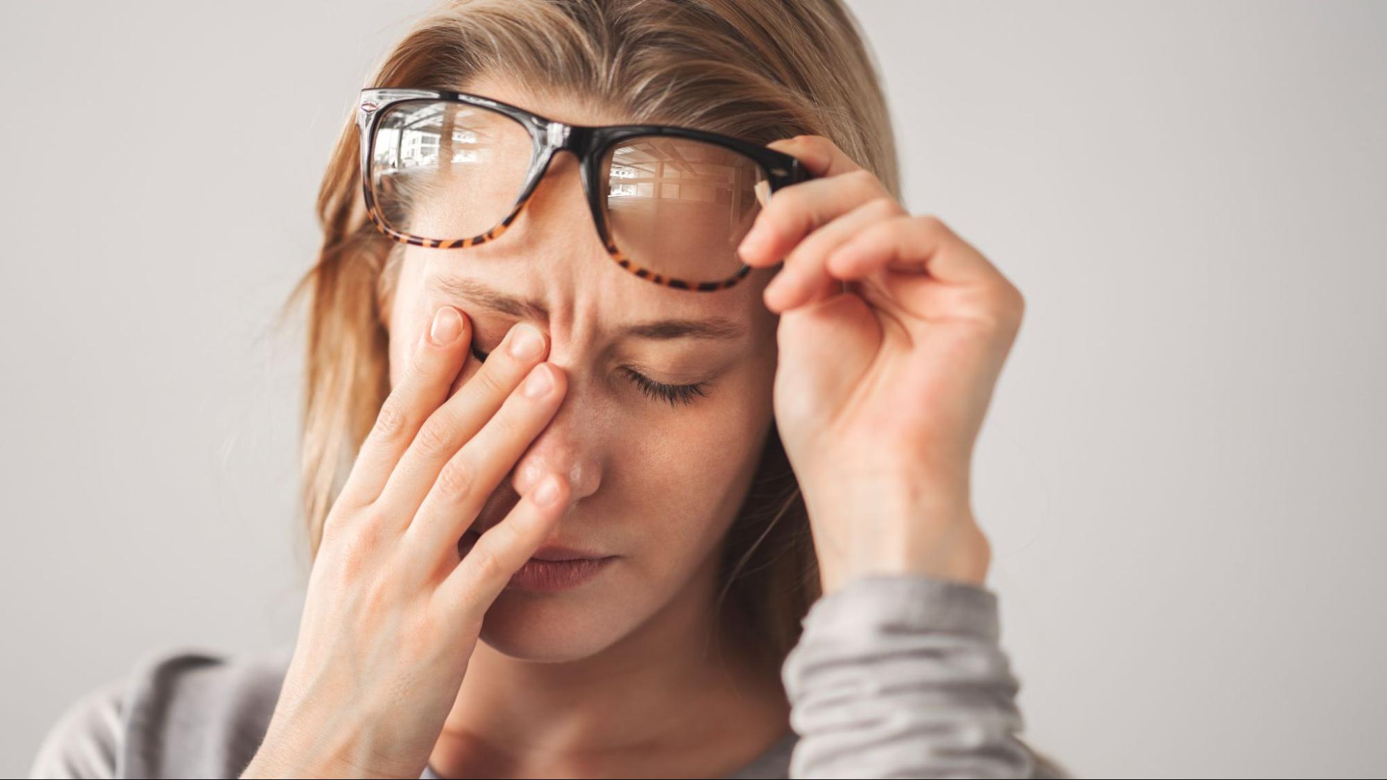
  • Avoid rubbing your eyes, which releases more histamine and makes itching worse

Contact Lenses and Eye Allergies

Wearing contact lenses during allergy season adds another layer of irritation. Airborne allergens stick to lens surfaces and sit directly against your eye. A few things that help:

  • Switch to daily disposable lenses during peak pollen season
  • Talk to your eye doctor about allergy-safe lens options
  • Follow your replacement schedule strictly
  • Consider glasses on high-symptom days

If contact lenses consistently make your eye symptoms worse, a proper contact lens exam can identify whether fit, lens material, or care routine is the issue.

woman rubbing eyes

Allergy Shots

For people with moderate to severe symptoms that don’t respond well to eye drops or oral medications, allergy shots (immunotherapy) can gradually reduce how strongly your immune system reacts to a specific allergen. This is a longer-term treatment managed through an allergist, but it can significantly reduce how much your symptoms affect daily life over time.

When Symptoms Point to Something More Serious

Most eye allergy cases are annoying but not dangerous. However, there are situations where you should see an eye doctor rather than waiting it out:

  • Symptoms that don’t improve with over-the-counter treatment
  • Eye pain or significant light sensitivity
  • Vision changes
  • Thick or unusual discharge, which may signal bacterial infections
  • Symptoms that look like allergic conjunctivitis but don’t follow a seasonal pattern

Atopic keratoconjunctivitis, for example, can cause corneal damage without proper treatment. This more severe form of eye allergy requires close monitoring by an eye doctor to prevent long-term complications. If your eye symptoms feel more intense than typical seasonal allergy flare-ups, an exam can rule out something that needs more targeted care.

The Bottom Line on Eye Allergy Relief

Eye allergy symptoms are common, but they don’t have to take over your daily life. The right approach depends on what’s triggering your reaction, how severe your symptoms are, and whether other conditions like dry eye are in the mix.

For most people, antihistamine eye drops and artificial tears offer real short-term relief. Managing allergen exposure at home and during pollen season helps reduce how often symptoms flare. For persistent or worsening cases, an eye doctor can pinpoint the cause and recommend proper treatment, whether that’s prescription antihistamine eye drops, adjusted contact lens care, or a referral for allergy shots.

At Visionary Eye Doctors, we see patients across Washington, DC and Maryland who are struggling to manage allergy-related eye symptoms, often alongside dry eye or other concerns. If itchy, watery eyes are getting in the way, we’re here to help you find lasting relief.

We also offer in-office allergy testing to help pinpoint exactly what’s triggering your symptoms, so your treatment plan is tailored to you. Book an appointment today and get answers from a team that genuinely knows eyes.

Senior man has cataract. Generally, cataracts are common in older people.

What Causes Cataracts? Understanding the Factors Behind Cloudy Vision

The Short Answer: A cataract forms when proteins in your eye’s lens break down and clump together, creating a cloudy area that blocks light rays from reaching the retina clearly. While aging is the most common cause, other factors like diabetes, eye injuries, and genetics can also lead to cataract formation.

More than half of Americans age 80 and older either have cataracts or have had cataract surgery, according to the National Eye Institute. Understanding what causes this common condition can help you take steps to protect your vision and know when to schedule an eye exam.

How Your Eye’s Lens Works

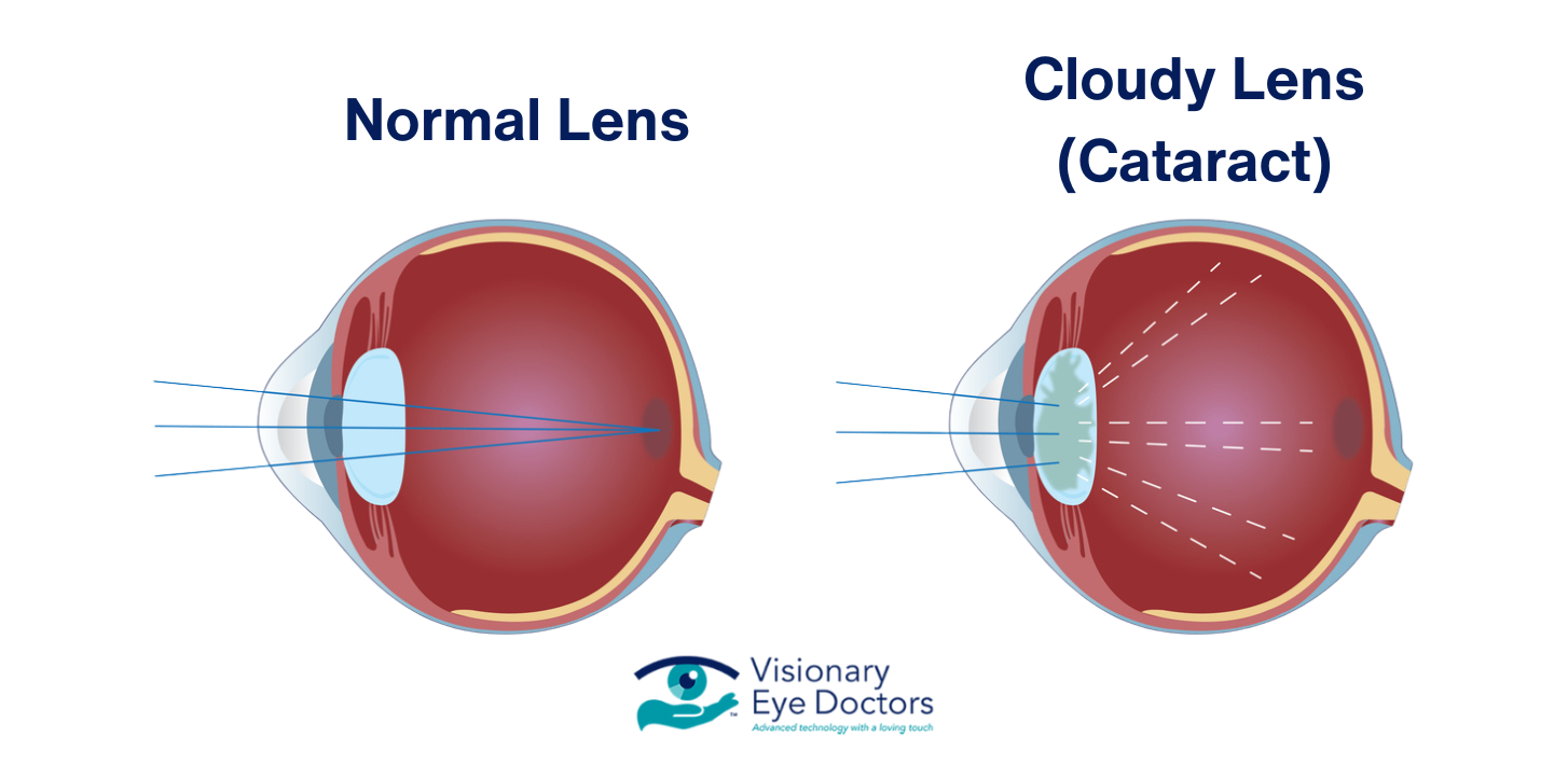
Your eye has a natural lens that sits behind your iris (the colored part of your eye). This lens focuses light rays onto the retina at the back of your eye, which sends signals through the optic nerve to your brain to create the images you see.

A healthy lens is clear, like a clean window. It is made mostly of water and proteins arranged in a precise pattern that allows light to pass through. When those proteins start to break down and clump together, they create a cloudy lens that scatters light instead of focusing it. That clouded lens is a cataract.

Normal lens vs cloudy lens (cataract)

The Main Causes of Cataracts

Aging

Age-related cataract is the most common type. Over time, the proteins in your eye lens begin to break down due to years of oxidative stress and exposure to ultraviolet light. This natural wear and tear causes the lens to gradually become less transparent.

Most people begin developing cataracts around age 40, though vision problems may not become noticeable until age 60 or later. Among older adults, cataracts are the leading cause of vision loss worldwide.

Medical Conditions

Several medical conditions can increase your risk of cataract formation:

  • Diabetes: High blood sugar levels can cause the eye lens to swell and change shape, speeding up protein breakdown. People with diabetes have a higher risk of developing cataracts at a younger age.
  • Autoimmune diseases: Conditions like rheumatoid arthritis and lupus (and the corticosteroid medications used to treat them) are linked to earlier cataract development.
  • Previous eye surgery or retinal detachment: Earlier procedures on the eye can increase the chances of a cataract forming later.

Any medical condition that affects blood flow or inflammation in the body can potentially contribute to changes in the lens over time. If you have a chronic health condition, let your eye doctor know so they can monitor your eyes more closely.

Eye Injuries

A traumatic cataract can develop after a blow, puncture, or chemical burn to the eye. The injury damages the structure of the lens capsule, allowing fluid to enter and disrupt the organized protein fibers. A traumatic cataract may appear soon after the injury or develop years later.

Even minor injuries that seem to heal fully can leave behind damage that leads to cataract formation down the road. Wearing protective eyewear during sports, yard work, and home improvement projects is one of the simplest ways to reduce this risk factor.

Genetics and Congenital Cataracts

Some babies are born with a congenital cataract or develop one in early childhood. These may be inherited or caused by infections during pregnancy, such as rubella. A congenital cataract can affect a child’s vision development, so early detection by a pediatric ophthalmologist is important. If the cataract is large enough to block normal vision, surgery may be needed to protect the child’s vision as they grow.

Family history also plays a role in age-related cataracts. If your parents or siblings developed cataracts early, you may be at higher risk for earlier onset as well.

Types of Cataracts

There are several types of cataracts, each affecting a different part of the lens:

  • Nuclear cataract: This is the most common age-related type. A nuclear cataract forms in the center (nucleus) of the lens. It often begins with a process called nuclear sclerosis, where the lens gradually hardens and yellows. Over time, it may cause blurry vision and difficulty distinguishing colors.
  • Cortical cataract: This type starts as white, wedge-shaped, cloudy patches on the outer edge of the lens and slowly extends toward the center. It can cause problems with glare and contrast.
  • Posterior subcapsular cataract: This type forms at the back of the lens, just inside the lens capsule. It tends to progress faster than other types and can cause difficulty reading, double vision, and glare from headlights at night. People who take corticosteroids or have diabetes are at higher risk for this type.

It is possible to have more than one type of cataract in the same eye. Your eye doctor can identify the specific type during a dilated eye exam, which helps guide the best approach for monitoring or treatment.

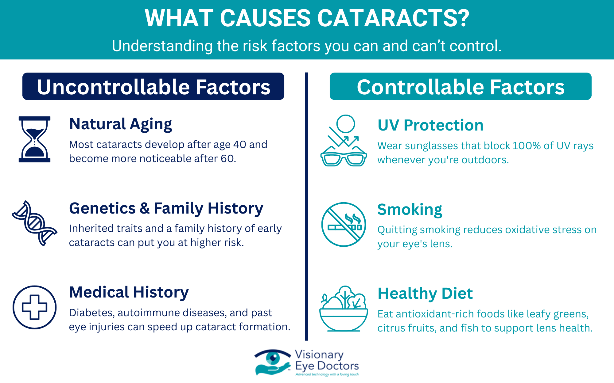
Risk Factors That Speed Up Cataract Formation

While you cannot fully prevent cataracts, being aware of the risk factors can help you slow their progression:

  • Prolonged UV exposure without sunglasses
  • Smoking and heavy alcohol use
  • Long-term use of corticosteroid medications
  • A family history of early cataracts
  • Previous eye injuries or eye surgery
  • Obesity
  • High blood pressure

Reducing these risk factors where possible can help. Wearing UV-blocking sunglasses, eating a healthy diet rich in antioxidants (like leafy greens, citrus fruits, and fish), managing conditions like diabetes, and quitting smoking are all practical steps you can take. Research from the American Academy of Ophthalmology supports the link between lifestyle choices and the pace of cataract development.

What causes cataracts? infographic

Cataract Symptoms to Watch For

Cataracts usually develop slowly. You might not notice changes at first, but over time, cataract symptoms can include:

  • Cloudy vision or blurry vision that worsens gradually
  • Colors appearing faded or yellowed
  • Increased sensitivity to glare from headlights or sunlight
  • Double vision in one eye
  • Needing brighter light for reading and other daily activities
  • Frequent changes to your eyeglass or contact lens prescription

These symptoms can overlap with other vision problems, which is why a professional evaluation matters. If any of these sound familiar, schedule an eye exam. Your eye doctor can check for cataracts and other conditions that may be affecting your normal vision.

How Cataracts Are Treated

In the early stages, updated eyeglasses or contact lenses may help you manage mild vision changes. But as the cataract grows and begins to interfere with daily activities like driving, reading, or working, cataract surgery becomes the most effective treatment.

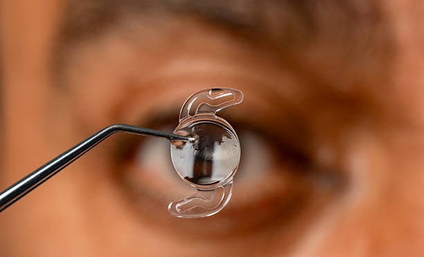
During cataract surgery, your surgeon removes the clouded lens and replaces it with an artificial lens called an intraocular lens (IOL). This artificial lens is permanent and designed to restore clear vision. There are several IOL options available depending on your lifestyle and visual needs.

Cataract surgery is one of the most commonly performed procedures in the United States, with a high success rate. Most patients notice a significant improvement in their vision within a few days of the procedure.

What About Posterior Capsular Opacification?

Some patients notice cloudy vision returning months or years after cataract surgery. This is called posterior capsular opacification (sometimes referred to as a “secondary cataract”). It happens when the thin membrane behind the intraocular lens becomes cloudy. A quick, painless laser procedure called a YAG capsulotomy can correct it, and the results are typically immediate.

Protect Your Vision with Regular Eye Exams

Cataracts are one of the most common causes of vision loss, but they are also one of the most treatable. Knowing what causes cataracts, from aging and oxidative stress to medical conditions and eye injuries, gives you the information you need to be proactive about your eye health.

At Visionary Eye Doctors, our team provides personalized cataract care for patients across Washington, DC, and Maryland. From early detection during a routine eye exam to advanced cataract surgery with premium intraocular lens options, we are here to help you see clearly at every stage.

Book your appointment today to get ahead of cataracts and protect the vision you depend on.

Early Signs of Glaucoma: What You Need to Know to Protect Your Vision

The short answer: The most common type of glaucoma, open-angle glaucoma, typically has no early warning signs or noticeable symptoms. This is why glaucoma is often called the “silent thief of sight.” However, acute angle-closure glaucoma can cause sudden, severe symptoms requiring immediate medical attention. Regular comprehensive eye exams are the only reliable way to detect glaucoma before permanent vision loss occurs.

What Is Glaucoma?

Glaucoma is a group of eye diseases that cause progressive damage to the optic nerve, the vital connection that sends visual information from your eyes to your brain. In most cases, this damage is associated with elevated intraocular pressure (the pressure inside your eye).

Your eye constantly produces a clear fluid called aqueous humor. This fluid nourishes the eye and maintains its shape. It normally drains through the trabecular meshwork at the drainage angle where the iris meets the cornea. When this drainage system doesn’t work properly, fluid builds up and increases pressure, potentially damaging the optic nerve over time.

If left untreated, glaucoma can lead to permanent vision loss, making it the second leading cause of blindness worldwide. However, with early detection and proper treatment, most people can preserve their vision for years to come.

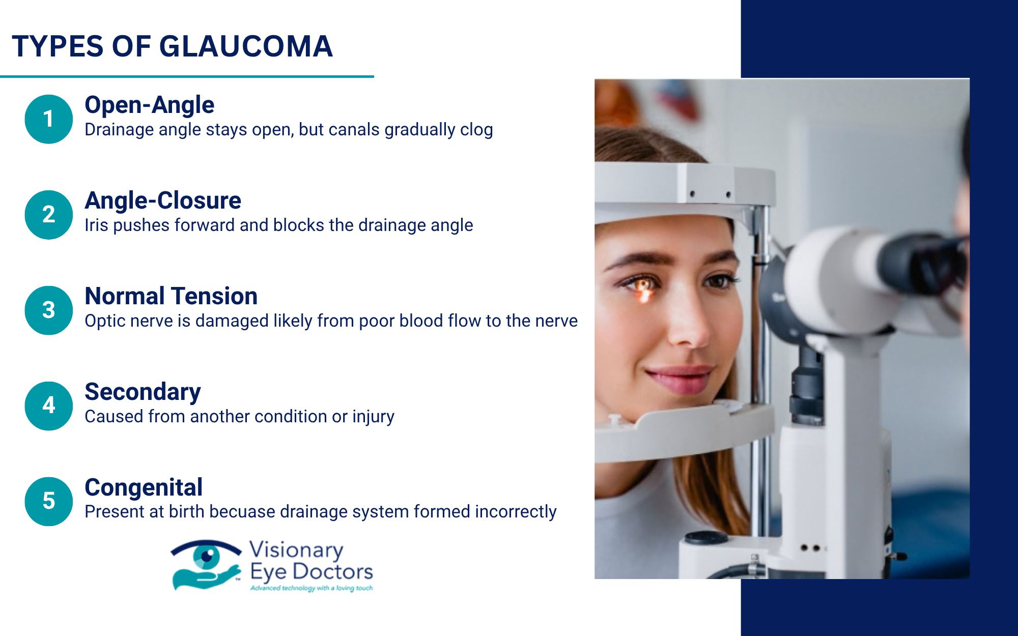
Types of Glaucoma

Open-Angle Glaucoma

Primary open-angle glaucoma is the most common form, accounting for about 90% of cases. The drainage angle remains open, but the trabecular meshwork becomes less efficient over time, causing a gradual increase in eye pressure.

This type develops so slowly that most people don’t notice changes until significant optic nerve damage has occurred. Vision loss typically begins with peripheral vision, which many people don’t immediately recognize. Because the changes happen gradually, your brain often compensates, making it even harder to detect without a professional exam.

Angle-Closure Glaucoma

Acute angle-closure glaucoma occurs when the iris bulges forward and completely blocks the drainage angle, causing a rapid spike in eye pressure. This is a medical emergency requiring immediate treatment to prevent permanent damage.

Chronic angle-closure glaucoma develops more gradually as the iris slowly blocks the drainage angle over time. Some people may experience intermittent episodes where the angle temporarily closes, causing brief symptoms that resolve on their own before a full acute attack occurs.

Normal-Tension Glaucoma

Some people develop optic nerve damage even with normal eye pressure. Researchers believe factors such as reduced blood flow to the optic nerve may play a role. This highlights why comprehensive eye exams evaluating optic nerve health are essential, not just pressure checks.

Early Signs and Symptoms of Glaucoma

Infographic: Early signs of glaucoma you might miss

Open-Angle Glaucoma: The Silent Threat

The most challenging aspect of open-angle glaucoma is that there are typically no obvious symptoms in the early stages. Up to half of the people with glaucoma don’t know they have it until the disease has progressed significantly.

As the disease progresses, you may notice:

  • Patchy blind spots in your peripheral vision
  • Difficulty seeing objects to your side while looking straight ahead
  • In advanced stages, tunnel vision occurs, where only central vision remains

Acute Angle-Closure Glaucoma: A Medical Emergency

Unlike open-angle glaucoma, acute angle-closure produces sudden, severe symptoms:

  • Severe eye pain and intense headache
  • Sudden blurry or decreased vision
  • Halos or rainbow-colored rings around lights
  • Eye redness, nausea, and vomiting

Subtle Warning Signs to Watch For

Some people may experience intermittent warning signs before a full acute attack or in the early stages of certain types of glaucoma. These can include occasional blurry vision, mild headaches or eye discomfort, seeing halos around lights at night, and needing more light to see clearly. If you notice any of these symptoms, schedule an appointment with your eye doctor for evaluation.

Who Is at Higher Risk?

While anyone can develop glaucoma, certain factors increase your risk:

  • Age: Risk increases significantly after 60. For African Americans, elevated risk begins around age 40.
  • Family history: Having an immediate family member with glaucoma increases your risk four to nine times.
  • Race and ethnicity: African Americans are six to eight times more likely to develop glaucoma. Hispanic Americans have an elevated risk after 60, and people of Asian descent have a higher risk for angle-closure glaucoma.
  • Medical conditions: Diabetes, high blood pressure, and heart disease can increase risk.
  • Eye factors: Previous eye injuries, severe nearsightedness, thin corneas, and elevated eye pressure.

How Eye Doctors Detect Glaucoma

A comprehensive eye exam includes several tests to evaluate your glaucoma risk and detect early signs of the disease. Since vision loss from glaucoma is permanent and cannot be reversed, these exams are critical for catching the condition before damage occurs.

  • Tonometry: Measures the pressure inside your eye
  • Ophthalmoscopy: Examines the shape and color of your optic nerve for signs of damage
  • Visual field testing: Maps your complete field of vision to detect blind spots you might not notice
  • Gonioscopy: Examines the drainage angle to determine your glaucoma type or risk
  • Pachymetry: Measures corneal thickness, which can affect the accuracy of pressure readings

These tests are painless and can identify problems before you experience any vision changes.

Glaucoma Treatment Options

While there’s no cure for glaucoma, effective treatments can control the disease and prevent further vision loss. The goal of treatment is to lower eye pressure to a level that prevents additional optic nerve damage.

Prescription eye drops are often the first line of treatment, working to reduce eye pressure by either decreasing fluid production or improving drainage. Laser treatment can improve fluid drainage: selective laser trabeculoplasty for open-angle glaucoma, or laser peripheral iridotomy for angle-closure glaucoma. Surgery may be recommended when other treatments aren’t sufficient, creating new drainage pathways to lower eye pressure.

Why Early Detection Matters

Vision loss from glaucoma is permanent and cannot be reversed, even with treatment. However, early detection and treatment can stop or significantly slow the progression of the disease, preserving your remaining vision for years or even decades.

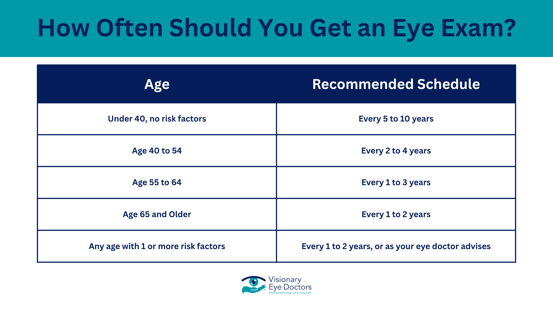
The American Academy of Ophthalmology recommends that adults have a comprehensive eye exam around age 40, when early signs of eye disease often begin. If you have risk factors such as family history, elevated eye pressure, or certain health conditions, your eye doctor may recommend more frequent exams to monitor for changes.

Steps to Protect Your Eye Health

Infographic: How to protect your vision from glaucoma

You can’t prevent glaucoma, but you can take proactive steps to catch it early and protect your sight.

  • Schedule regular eye exams starting at age 40, with more frequent visits if you have risk factors.
  • Know your family history and share it with your eye doctor.
  • Manage your overall health by controlling diabetes and blood pressure, which supports good eye health.
  • Protect your eyes from injury by wearing protective eyewear during sports and activities.
  • Follow your treatment plan consistently if you’ve been diagnosed with glaucoma, even if you feel fine.

Schedule Your Eye Exam at Visionary Eye Doctors

Glaucoma may be the “silent thief of sight,” but it doesn’t have to steal your vision. Early detection through regular comprehensive eye exams is your best defense against this sight-threatening condition.

If you have risk factors for glaucoma or haven’t had an eye exam recently, don’t wait. Schedule a comprehensive eye exam with the experienced team at Visionary Eye Doctors. Our advanced diagnostic technology allows us to detect glaucoma in its earliest stages, giving you the best chance to preserve your vision.

With early detection and proper treatment, most people with glaucoma can maintain their sight and continue enjoying the activities that matter most. Taking that first step to schedule an exam could be the most important thing you do for your long-term eye health.

Man scratching his eye

How Can You Tell if You Scratched Your Cornea?

The Short Answer: The main signs to watch for if you’ve scratched your cornea include eye pain, a gritty sensation like something is stuck in your eye, light sensitivity, excessive tearing, and blurry vision. A scratched cornea, medically known as a corneal abrasion, often causes discomfort that worsens with blinking. While minor abrasions may heal within a few days, it’s essential to see an eye doctor to avoid complications such as infections or corneal ulcers.

What Is a Corneal Abrasion?

A corneal abrasion is a scratch on the surface of your cornea, which is the clear, dome-shaped tissue covering the front of your eye. The cornea is highly sensitive, containing more nerve endings than almost any other body part, so even a small scratch can be intensely painful.

Corneal abrasions range from minor injuries that heal quickly to deeper abrasions that require medical attention. The severity depends on the scratch’s depth and whether any foreign material is embedded in the eye.

Common Causes of a Scratched Cornea

Understanding how corneal injuries happen can help prevent them. The most common causes of corneal scratches include:

  • Foreign objects in the eye: Dust, sand, metal shavings, and other small particles can scratch the cornea if they get trapped under your eyelid or come in direct contact with the eye.
  • Contact lens problems: Wearing contact lenses too long, sleeping in contacts not designed for overnight wear, or inserting damaged lenses can cause a corneal scratch. Poor lens hygiene is also a major contributor to corneal abrasions.
  • Eye rubbing: Rubbing your eyes vigorously, especially when a foreign object is present, can drag debris across the cornea, causing injury.
  • Accidental pokes: Fingernails, makeup applicators, or other objects near the eye can accidentally scratch the cornea.
  • Sports and recreation: Sports equipment, branches from hiking, or flying debris during outdoor activities can cause corneal injuries, especially if eye protection isn’t worn.
  • Workplace hazards: Jobs involving metalwork, woodworking, or environments with airborne particles increase the risk of corneal abrasions if protective eyewear isn’t used.

Signs and Symptoms of a Corneal Scratch

So, how can you tell if you’ve scratched your cornea? Here are the most common symptoms:

  • Eye pain: Sharp, immediate pain in the affected eye is usually the first sign. The discomfort can range from mild irritation to intense pain, depending on the severity of the scratch.
  • Foreign body sensation: You may feel like something is stuck in your eye, even after flushing it with water or saline.
  • Light sensitivity: Bright lights may cause discomfort or pain in the scratched eye. This can make it difficult to be in well-lit rooms or outdoors.
  • Excessive tearing: Your eye may produce more tears as it tries to flush out irritants and protect the damaged area.
  • Blurry vision: The scratch can temporarily blur your vision, particularly if it’s in the center of your cornea.
  • Redness: The white part of your eye may appear red or bloodshot due to inflammation and irritation.
  • Pain with blinking: Blinking can worsen the discomfort since your eyelid moves over the damaged area.
  • Headache: Some people may experience headaches from the strain caused by eye pain and light sensitivity.

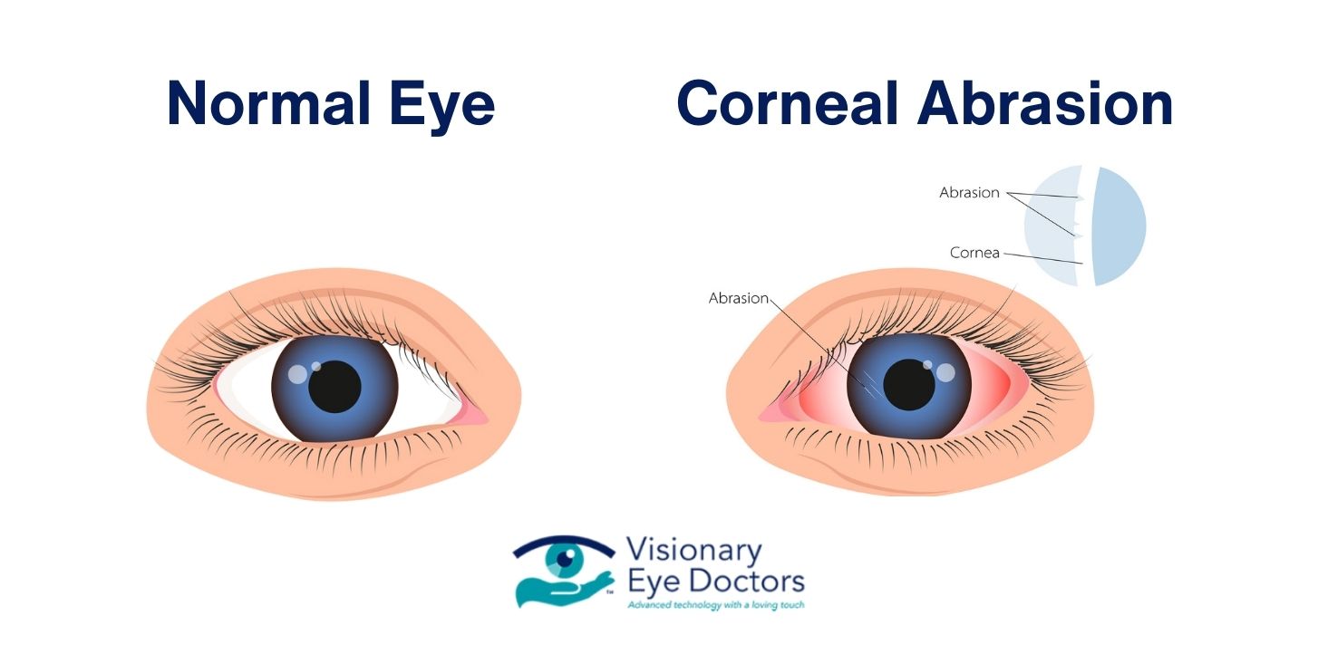
Normal Eye vs Corneal Abrasion

Minor Abrasion vs. Serious Corneal Injury

A minor scratch may cause mild to moderate discomfort and typically heals within a few days. However, larger abrasions or deeper injuries can lead to severe pain, vision problems, and may require medical treatment to avoid complications. If symptoms are intense or don’t improve within 24 hours, it’s likely a more serious injury.

What to Do If You Think You Scratched Your Cornea

If you think you’ve scratched your cornea, here’s what to do:

  1. Don’t rub your eye: Rubbing your eye can push a foreign object deeper or worsen an existing scratch.
  2. Rinse gently: Flush the eye with clean water or saline to remove debris. Tilt your head so the affected eye is down and let the water wash over it.
  3. Blink frequently: Blinking can help flush out small particles and spread your tears across the eye.
  4. Lift the eyelid gently: If debris is trapped under your eyelid, gently pull the lid away from your eye and look in different directions to dislodge it.
  5. Remove contact lenses: Take out your contact lenses immediately and avoid putting them back in until you’ve seen an eye doctor.
  6. Protect your eye: If light sensitivity is severe, wear sunglasses and avoid touching or pressing on your eye.

What Not to Do

Don’t use tweezers or cotton swabs to remove anything from your eye, since that can make the injury worse. Avoid eye drops unless your healthcare provider prescribed them, and don’t patch your eye tightly unless your doctor tells you to.

When to See an Eye Doctor

You should consult an eye doctor if:

  • Pain persists after rinsing
  • Blurry vision or vision problems become significant
  • Symptoms worsen over time
  • You see a foreign object embedded in your eye
  • The injury was caused by a high-speed object, metal or a chemical
  • You notice signs of infection, such as discharge or increasing redness
  • The injury involves a child’s eye

For serious injuries or if you can’t reach your doctor, head to the emergency room. Untreated corneal scratches can develop into infections like corneal ulcers, which can lead to permanent vision loss.

How Eye Doctors Diagnose a Corneal Abrasion

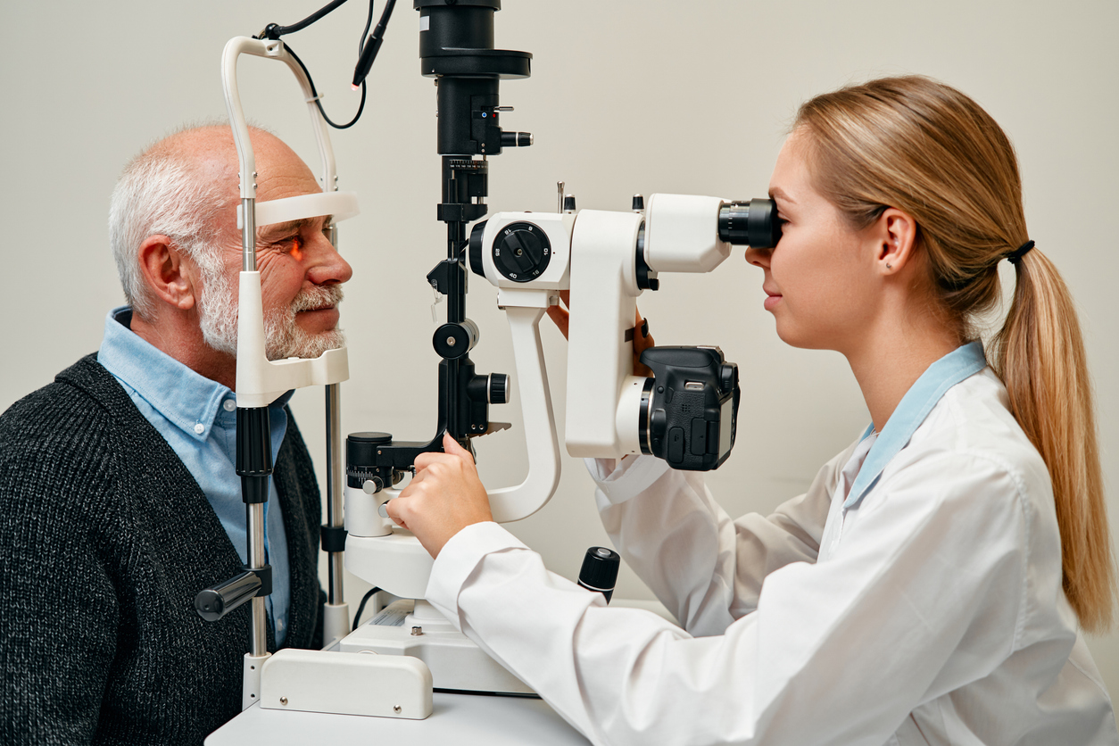
An eye doctor will use a slit lamp to examine your eye under magnification and light. They may apply fluorescein dye, which turns green under blue light, helping them see even tiny abrasions. The doctor will check for any foreign particles, assess the depth of the injury, and examine for signs of infection to determine the best course of treatment.

Treatment Options for Corneal Abrasions

Treatment depends on the severity of the scratch:

  • Antibiotic eye drops/ointment: These help prevent infection while the cornea heals.
  • Pain management: Over-the-counter pain relievers can manage discomfort. Prescription eye drops may also be provided.
  • Bandage contact lens: For larger abrasions, a special lens may be used to reduce pain and protect the eye during healing.
  • Lubricating eye drops: These keep the eye moist and comfortable as it heals.

The Healing Process

Most minor corneal abrasions heal within 24-72 hours. Larger abrasions may take a few days to a week to heal completely. Be sure to follow your doctor’s instructions and attend any follow-up appointments to monitor healing.

Corneal Abrasion vs. Pink Eye

Although both conditions can cause redness and irritation, pink eye (conjunctivitis) is typically accompanied by itching and discharge, while a corneal abrasion results in sharper pain, light sensitivity, and a history of trauma to the eye. If you’re unsure, see your eye doctor for a diagnosis.

Preventing Corneal Scratches

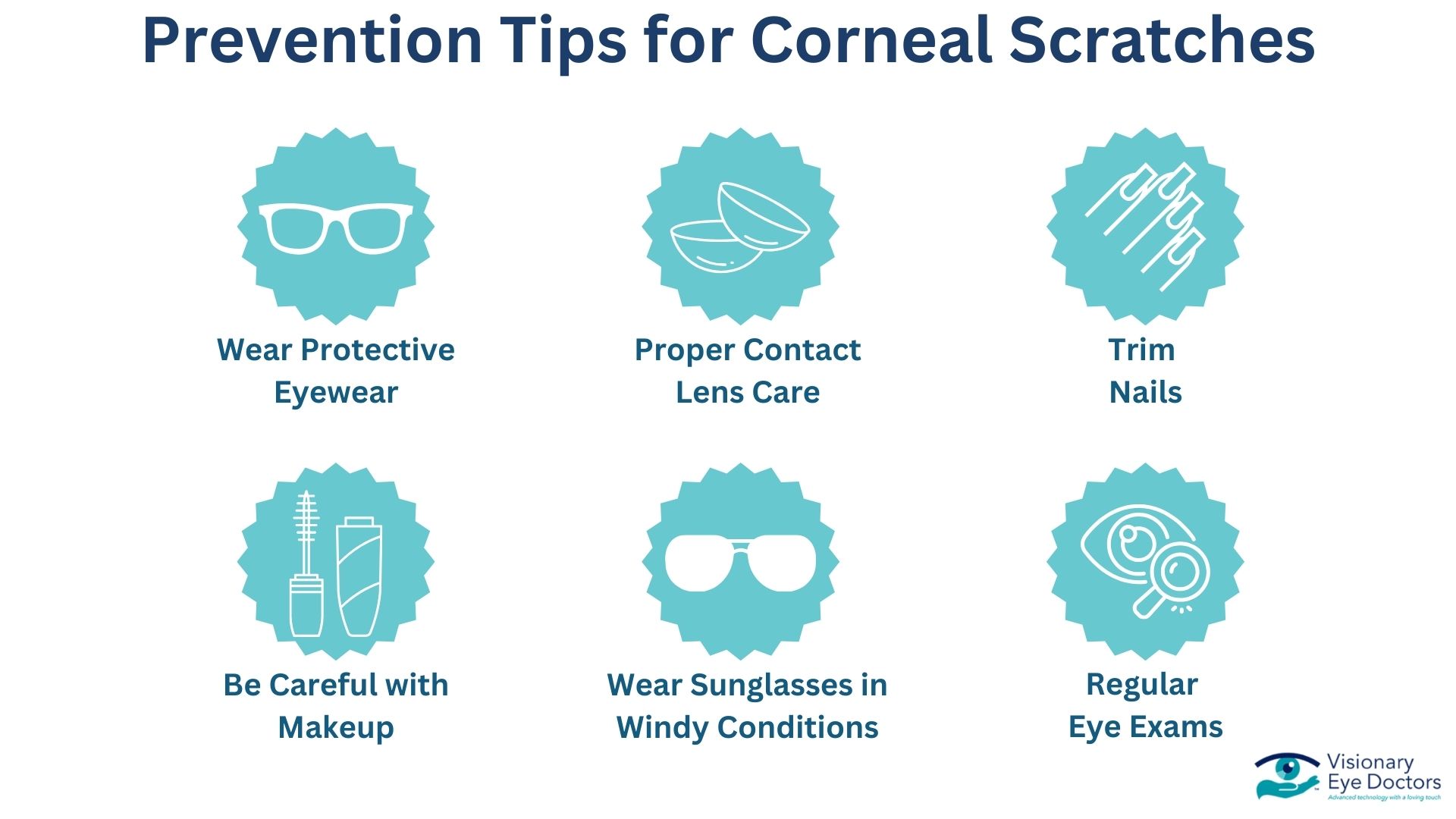
Prevention Tips for Corneal Scratches

You can reduce the risk of corneal abrasions by taking the following precautions:

  • Wear protective eyewear for work and sports
  • Practice proper contact lens care
  • Keep nails trimmed to prevent accidental scratches
  • Be careful with eye makeup and avoid sharing cosmetics
  • Wear sunglasses in windy conditions
  • Schedule regular eye exams to catch any issues early

Protect Your Vision with Expert Care at Visionary Eye Doctors

A scratched cornea can be painful, but with the right treatment, most abrasions heal fully. Recognizing symptoms early and seeking professional care is key to preventing complications and maintaining healthy vision. If you’re experiencing symptoms of a corneal scratch, don’t wait. Schedule an appointment with Visionary Eye Doctors today for the expert care your eyes deserve. For more corneal information, visit our Corneal Care Center page.

Female optician doing eye test with eye chart on her patient in ophthalmology clinic

Can Astigmatism Be Corrected?

The short answer: Yes, astigmatism can be corrected with glasses, contact lenses, or surgery. Many effective treatments are available to help you achieve clear vision, whether you have mild or severe astigmatism.

Astigmatism is a common refractive error that happens when your cornea or lens has an irregular shape. Whether you have mild astigmatism or more severe astigmatism, understanding your options for vision correction is the first step toward clearer vision.

Astigmatism affects millions of people worldwide. Some people have mild symptoms that barely bother them, while others have severe astigmatism that makes daily activities challenging. Here’s the good news: astigmatism can absolutely be corrected, and today’s treatments work really well.

Understanding Astigmatism: The Basics Made Simple

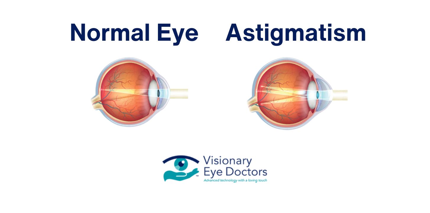
Astigmatism happens when your eye’s cornea or lens isn’t perfectly curved. Instead of being perfectly round like a basketball, an irregularly shaped cornea is more like a football. This irregular shape stops light from focusing properly on your retina, which causes blurred vision at all distances.

Most people are actually born with some degree of astigmatism – it’s incredibly common. The condition can also develop later in life due to eye injuries, certain eye diseases, or even after eye surgery.

infographic comparing a normal eye to astigmatism

There are two main types you should know about:

  • Regular astigmatism: The most common type, where the cornea curves more in one direction than the other
  • Irregular astigmatism: Less common and usually happens because of an eye injury or certain eye conditions

Common symptoms of astigmatism include:

  • Blurry vision at all distances
  • Eye strain and fatigue
  • Headaches
  • Trouble seeing clearly both up close and far away
  • Squinting to try to see better

Unlike other vision problems that only affect distance vision or near vision, astigmatism usually causes blurred vision no matter what you’re looking at. Corneal astigmatism occurs when the cornea has an irregular shape, while lenticular astigmatism happens when the eye’s natural lens is irregularly shaped. Some people also have myopic astigmatism, which combines nearsightedness with astigmatism.

Glasses and Contact Lenses: The Go-To Solutions

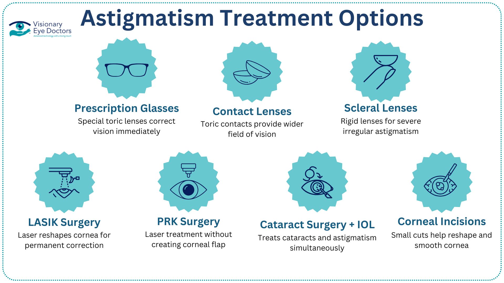
astigmatism treatment options

Prescription Glasses

Prescription eyeglasses are still the most popular way to correct astigmatism. These glasses use special toric lenses that have different powers in different areas to make up for your eye’s irregular shape. Your eye doctor will figure out exactly what prescription you need during a comprehensive eye exam.

Contact Lenses

Toric contact lenses are specially made corrective lenses for people with astigmatism. Unlike regular contact lenses, toric contact lenses are designed with varying powers across different areas of the lens to correct astigmatism. They also have a special weight distribution that keeps them properly aligned in your eye. Many people prefer contacts because they provide a wider field of vision and won’t fog up in changing weather conditions.

Both prescription lens options work great for most people with astigmatism. Your eye doctor can help you decide whether glasses or contacts work better for your lifestyle and your specific type of astigmatism.

LASIK and Laser Surgery: Permanent Solutions

LASIK Surgery

LASIK surgery (laser in situ keratomileusis) is the most common refractive surgery for astigmatism correction. During LASIK, a surgeon uses a femtosecond laser to create a thin flap in your cornea, then reshapes the tissue underneath to fix the irregular shape that’s causing your vision problems.

PRK Surgery

Photorefractive keratectomy (PRK) is another laser vision correction option. It’s similar to LASIK surgery, but PRK doesn’t require creating a corneal flap, which makes it a good choice for people with thinner corneas.

Most people who get laser surgery see major improvements in their vision. However, some might have residual astigmatism that needs additional treatment or you might still need corrective lenses for the best visual outcome. Good candidates for refractive surgery usually have stable vision, healthy eyes, and realistic expectations about what the surgery can do. Your eye doctor will do thorough testing to see if you’re a good candidate for these procedures.

Other Treatment Options: More Ways to Get Clear Vision

Scleral Lenses

Scleral lenses are large, rigid contact lenses that vault over your entire cornea. These specialized lenses work really well for people with irregular astigmatism or severe corneal irregularities that can’t be fixed with regular toric contact lenses.

Cataract Surgery with Toric IOL

For people who are developing cataracts along with astigmatism, cataract surgery with toric IOL (intraocular lens) implantation can fix both problems at the same time. During this procedure, a cataract surgeon removes the cloudy natural lens and replaces it with a toric intraocular lens that’s designed to correct astigmatism.

Corneal Relaxing Incisions

Corneal relaxing incisions are small, precise cuts made in the cornea during cataract surgery or as a standalone procedure. These incisions help reshape the cornea and reduce astigmatism.

Some eye doctors might recommend combining treatments for the best results. For example, you might have cataract surgery with intraocular lenses and still need glasses to fine-tune your vision.

Choosing the Right Treatment for Your Life

Several factors affect which astigmatism correction method works best for you:

  • Cost considerations: While glasses and contact lenses cost money over time, laser surgery and other eye surgery options cost more upfront but might save you money in the long run
  • Your health factors: Your age, overall eye health, and how severe your astigmatism is all matter when choosing treatment. People with mild astigmatism might do great with glasses or contacts, while those with more complex vision problems might benefit from surgical options
  • Lifestyle needs: If you’re active in sports, you might prefer contact lenses or laser surgery for outdoor activities. People who work a lot on computers might find certain treatments reduce eye strain better than others

Make sure to ask your eye doctor about recovery time, potential risks, and what kind of visual outcomes you can expect with each option. Understanding what to expect helps you make the best choice for your situation.

Take the Next Step

Astigmatism is highly treatable, and you have many excellent options for correction. Modern vision correction technology offers solutions for every type of astigmatism, from mild cases to more complex irregular astigmatism or myopic astigmatism. Whether you prefer prescription eyeglasses, contact lenses, or advanced laser surgery techniques, there’s likely a treatment that fits your lifestyle and gives you the clearer vision you want.

The most important thing you can do is schedule a comprehensive eye exam with a qualified eye doctor. They’ll check your specific type of astigmatism, talk about your goals and lifestyle, and recommend the best treatment approach for your individual needs. With the right treatment, astigmatism doesn’t have to limit what you do or affect your quality of life.

At Visionary Eye Doctors, our experienced team knows that every patient’s vision needs are different. We offer the full range of astigmatism correction options, from the latest toric contact lenses to advanced refractive surgery techniques. Don’t let astigmatism keep affecting your daily life – schedule your consultation today and take the first step toward the clear vision you deserve. Clear vision is totally achievable, and we’re here to help you get there at our convenient locations in Washington, D.C., Rockville, Shady Grove, Gaithersburg, Bethesda, and Damascus, Maryland.

Botox® vs Fillers: Which Treatment is Right for You?

The Short Answer: Botox® works by relaxing facial muscles to reduce dynamic wrinkles caused by facial expressions, while dermal fillers add volume to smooth static wrinkles and restore facial features. Botox contains botulinum toxin that temporarily blocks nerve signals to muscles, whereas fillers use gel-like substances such as hyaluronic acid to plump and fill areas of volume loss. 

Both are effective cosmetic treatments, but they work in different ways to achieve a revitalized appearance. Understanding the difference between these injectable treatments helps you choose the right option for your aesthetic goals.

At Visionary Eye Doctors, Dr. Mahan, a Botox expert, specializes in advanced techniques and is well-known for training other physicians in these methods, including the Botox brow lift, which elevates the brows and makes the eyes appear more open and refreshed. Her expertise ensures that you receive the best care possible to achieve natural, lasting results.


Table infographic comparing Botox to fillers.

Botox

What Is Botox?

Botox is an injectable treatment derived from bacterium clostridium botulinum. It’s a cosmetic procedure that temporarily relaxes facial muscles to reduce the appearance of wrinkles caused by repeated muscle movement.

When you make facial expressions like smiling, frowning, or squinting, your facial muscles contract and create lines on your skin. Over time, these dynamic wrinkles become more pronounced.

How Does Botox Treatment Work?

Botox injections work by blocking nerve signals to specific facial muscles. When interrupted, the targeted muscles can’t contract as forcefully. This muscle relaxation smooths out the skin above, reducing the appearance of wrinkles and fine lines.

The effects of Botox typically appear within a few days and last three to six months.

Common Treatment Areas for Botox

Botox is most effective for treating dynamic wrinkles in the upper face:

  • Forehead lines: Horizontal lines across the forehead caused by raising your eyebrows
  • Frown lines: Vertical lines between the eyebrows
  • Crow’s feet: Fine lines that radiate from the outer corners of your eyes
  • Forehead wrinkles: Expression lines that deepen with repeated facial movements

Botox can also be used for the lower face: marionette lines (downturned mouth), TMJ/masseter (good for jaw clenching and also slimming of the jaw line), chin dimpling, “lip flip” 

Dr. Mahan also performs botox injections for medical conditions including TMJ dysfunction, facial spasms, blepharospasm, myokymia, and migraine. Some of these treatments may be covered by insurance. 

What to Expect During Treatment

A botox injection session typically takes just 10-15 minutes. Your healthcare provider will use a fine needle to inject small amounts of botulinum toxin into specific facial muscles. Most people describe the sensation as a slight pinch at the injection site. No anesthesia is usually needed, and you can return to normal activities immediately.

Potential Side Effects

People can experience minimal side effects from Botox treatment:

  • Mild bruising or swelling at the injection site
  • Headache (usually resolves within 24-48 hours)
  • Temporary drooping of the eyelid (rare)
  • Flu-like symptoms (uncommon)

Serious side effects are rare when administered by a board-certified surgeon. 

Dermal Fillers

What Are Dermal Fillers?

Dermal fillers are gel-like substances injected beneath the skin to restore lost volume, smooth wrinkles, and enhance facial features. Unlike Botox, which works on muscle movement, facial fillers physically fill in areas where volume has decreased due to aging.

Dermal filler injections can plump thin lips, soften facial creases, and improve the appearance of static wrinkles.

Types of Fillers

Different types of fillers serve different purposes:

  • Hyaluronic acid fillers: The most popular type. Hyaluronic acid is a naturally occurring substance in your body that helps skin retain moisture. These provide natural-looking results and can be reversed if needed. Dr. Mahan exclusively uses hyaluronic acid fillers due to their excellent safety profile and reversibility.
  • Calcium hydroxylapatite: A heavier filler used for deeper wrinkles and facial contouring.
  • Poly-l-lactic acid: Stimulates your skin to produce more collagen gradually, with results lasting up to two years.

Common Treatment Areas for Fillers

Dermal filler injections are most effective for static wrinkles and volume loss:

  • Smile lines: Lines running from the sides of your nose to the corners of your mouth
  • Marionette lines: Lines extending downward from the corners of your mouth
  • Lips: Add volume and define the lip border
  • Cheeks: Restore volume loss for a more youthful appearance
  • Under-eye area: Fill hollow areas to reduce a tired appearance
  • Temple: fill hollow temples that contribute to aged appearance

What to Expect During Treatment

A dermal filler injection session typically takes 30-60 minutes. Your plastic surgeon or dermatologist may apply numbing cream before injections. Many modern fillers also contain lidocaine for added comfort during the cosmetic treatment.

Results are visible immediately, though some swelling may temporarily mask the final outcome.

Potential Side Effects

Most side effects from dermal filler injections are mild and temporary:

  • Swelling and bruising at the injection site
  • Redness or tenderness in treated areas
  • Lumps or bumps under the skin (usually temporary)
  • Allergic reaction (rare, especially with hyaluronic acid fillers)

Serious complications are uncommon when performed by an experienced healthcare provider. Your surgeon will discuss these risks with you at the time of your consultation.

Infographic comparing how Botox and fillers work.

Botox vs Fillers: Key Differences

How They Work

Botox temporarily weakens  facial muscles to prevent dynamic wrinkles caused by facial movements. It doesn’t add volume but prevents wrinkle formation through muscle relaxation.

Fillers physically fill spaces beneath the skin to add volume and smooth static wrinkles. They don’t affect muscle movement but restore fullness to areas with volume loss.

Treatment Areas

Botox is most effective for the upper face, including forehead lines, frown lines, and crow’s feet caused by repeated facial expressions.

Fillers work best for the mid and lower face, such as smile lines, marionette lines, lips, and cheeks where volume restoration is needed.

Duration of Results

Botox effects typically last three to six months. Regular botox treatment maintains results.

Filler duration varies by type. Hyaluronic acid fillers last six to twelve months, while poly-l-lactic acid can last up to two years.

Can You Use Both Treatments?

Yes! Many people combine Botox and fillers in different ways to achieve complete facial rejuvenation:

  • Botox for forehead wrinkles and crow’s feet
  • Fillers for smile lines and volume loss in cheeks
  • A customized treatment plan addressing multiple concerns

Combining these cosmetic treatments creates balanced, natural-looking results that address both dynamic and static wrinkles.

Choosing the Right Treatment for Your Goals

Infographic on choosing the right treatment between Botox, fillers, or both.

Choose Botox if you want to:

  • Reduce wrinkles caused by facial expressions
  • Prevent new fine lines from forming
  • Smooth forehead lines, frown lines, or crow’s feet

Choose fillers if you want to:

  • Restore volume to areas of your face
  • Smooth wrinkles visible even at rest
  • Enhance lips or cheeks
  • Fill deep creases like smile lines or marionette lines

Consider both treatments if you want:

  • Balanced facial rejuvenation
  • To address concerns in both upper and lower face
  • Maximum improvement in your healthier-looking skin

Finding the Right Provider

Choosing a qualified healthcare provider is important for safe, effective results. Look for a board-certified dermatologist or plastic surgeon with extensive experience in injectable treatments.

At Visionary Eye Doctors, our oculofacial plastic surgery team specializes in treatments for the delicate eye area and upper face. We understand the unique anatomy around the eyes and can help you achieve natural-looking results while prioritizing your safety and satisfaction.

Your Path to a More Youthfull Appearance

Portrait of attractive brunette long-haired woman smiling at camera.

Both Botox and dermal fillers are proven, effective cosmetic treatments that can help you achieve your aesthetic goals. Whether you’re concerned about dynamic wrinkles from facial expressions or static wrinkles from volume loss, these injectable treatments offer safe solutions to smooth wrinkles and restore facial features.

The key is getting an accurate assessment from a qualified healthcare provider who can recommend the right cosmetic procedure for your individual needs. Some people benefit from Botox alone, others from fillers alone, and many find that combining both treatments provides the most comprehensive results.

Don’t let concerns about facial wrinkles or fine lines affect your confidence. Modern injectable treatments, administered by experienced professionals, help you maintain a natural, youthful appearance while preserving your unique facial movements.

Schedule a consultation with the oculofacial plastic surgery specialists at Visionary Eye Doctors to discuss whether Botox, fillers, or a combination of both is right for you. With the right cosmetic treatment plan, you can achieve the refreshed, radiant look you desire.

vision exam

Nearsightedness vs Farsightedness: Your Complete Guide to Common Vision Problems

The quick answer: Nearsightedness (myopia) means you see close objects clearly, but distant objects appear blurry. Farsightedness (hyperopia) means you see distant objects clearly, but close-up objects are blurry. Both are types of refractive error that affect how light rays focus in your eyes, and both are easily treatable with the right vision correction.

myopia hyperopia

Understanding the difference between these conditions helps you recognize symptoms and seek proper treatment from an eye doctor. Let’s explore everything you need to know about these vision problems.

Nearsightedness (Myopia): Everything You Need to Know

What Is Nearsightedness?

Nearsightedness, or myopia, is a common refractive error where nearby objects appear clear, but distant objects look blurry. This happens because your eye’s shape causes light rays to focus in front of the retina instead of directly on it. The retina is a light-sensitive tissue at the back of your eye that sends electrical impulses through the optic nerve to your brain.

What Causes Myopia?

Nearsightedness occurs when your eyeball is too long or your cornea (the clear front surface of your eye) is too curved. This changes where the focal point lands in your eye. Instead of light focusing directly on the retina for normal vision, it focuses in front of it, creating blurry vision when looking at distant objects.

Common Signs of Nearsightedness:

  • Squinting to see distant objects clearly
  • Difficulty reading road signs while driving
  • Sitting close to the TV or movie screen
  • Holding books or devices close to your face
  • Eye strain and headaches from trying to focus
  • Difficulty seeing the board at school or work

How Myopia Affects Daily Activities

Nearsighted individuals often struggle with activities that require distance vision:

  • Driving: Difficult, especially at night when road signs are harder to see
  • Playing Sports: Difficult when you can’t clearly see the ball or other players across the field
  • Entertainment: Watching movies in theaters becomes frustrating without proper vision correction
  • Social Situations: Recognizing faces across a room can be challenging

Treatment Options for Nearsightedness

Several effective treatments can restore clear vision for people with myopia:

  • Prescription Glasses: The most common solution, glasses with the correct prescription provide immediate, clearer vision for distant objects.
  • Contact Lenses: Contacts offer freedom from glasses and a wider field of vision. Daily, weekly, and monthly options are available.
  • Refractive Surgery: LASIK (laser in situ keratomileusis) and other laser procedures can permanently correct myopia by reshaping the cornea.
  • Orthokeratology (Ortho-K): Special contact lenses worn overnight temporarily reshape the cornea, providing clear vision during the day without glasses or contacts. Visionary Eye Doctors offers Ortho-K treatment for qualified patients.

When Does Myopia Develop?

Nearsightedness often starts in childhood and may worsen during the teenage years as the eyes continue growing. Environmental factors like increased screen time and less outdoor activity may contribute to its development. Regular eye exams are essential for monitoring a child’s vision and catching changes early.

Farsightedness (Hyperopia): Everything You Need to Know

What Is Farsightedness?

Farsightedness, or hyperopia, is a refractive error where distant objects appear clearer than close-up objects. Unlike myopia, hyperopia causes light rays to focus behind the retina instead of directly on it. This makes close-up vision more challenging than distance vision.

What Causes Hyperopia?

Farsightedness happens when your eyeball is too short or your cornea is too flat. This changes how light enters your eye and where it focuses. The focal point lies behind the retina, making nearby objects appear blurry while distant objects may remain relatively clear.

Common Signs of Farsightedness:

  • Difficulty reading books, newspapers, or phone screens
  • Eye strain during close work
  • Headaches after reading or computer work
  • Blurry vision when looking at close-up objects
  • Fatigue after activities requiring near vision
  • Children may have trouble with schoolwork or reading

How Hyperopia Affects Daily Activities

Farsighted vision significantly impacts activities requiring close-up vision:

  • Reading: Becomes uncomfortable and tiring without proper correction
  • Computer Work and Smartphones: Often cause eye strain and headaches during extended use
  • Hobbies: Crafts, sewing, or detailed work become difficult to perform
  • Daily Tasks: Simple activities like checking price tags while shopping can be challenging without proper corrective lenses

Treatment Options for Farsightedness

Multiple treatment options can help people with hyperopia achieve clear vision:

  • Reading Glasses: Over-the-counter or prescription reading glasses help with close-up tasks.
  • Bifocals and Progressive Lenses: These prescription glasses correct both distance and near vision in one lens.
  • Contact Lenses: Multifocal contacts or single-vision contacts can correct farsighted vision.
  • Refractive Surgery: Laser procedures can reshape the cornea to improve the eye’s ability to focus light properly on the retina.

When Does Hyperopia Develop?

Many people are born with mild farsightedness that may not cause problems until later in life. As we age, the eye’s natural lens becomes less flexible, making it harder to focus on close objects. This is why farsightedness often becomes noticeable in early adulthood or middle age, even if it was present from birth.

nearsighted vs farsighted signs

How Eye Doctors Diagnose Vision Problems

Getting an accurate diagnosis is the first step toward a clear vision. During a regular eye exam, your eye doctor will perform several tests to determine if you have a refractive error and what type of vision correction you need.

  • Visual Acuity Test: You’ll read letters on an eye chart to measure how well you see at various distances. This helps determine your level of visual acuity.
  • Refraction Test: Your eye doctor will use a device called a phoropter to test different prescription lenses. You’ll look through various lenses and tell the doctor which ones make your vision clearer.
  • Eye Health Examination: Beyond checking your vision, the eye doctor will examine the overall health of your eyes, including the retina and optic nerve.

The American Optometric Association recommends regular eye exams even if you don’t notice vision problems. Many refractive errors develop gradually, and early detection leads to better treatment outcomes.

Why Professional Diagnosis Matters

Online vision tests and self-diagnosis can’t replace a proper eye exam. Only a qualified eye doctor can determine the exact type and degree of your refractive error, rule out other eye health issues, and prescribe the correct treatment. They can also detect other vision problems that might be affecting your sight.

General Eye Health Tips

Taking care of your eyes goes beyond correcting refractive errors. These simple habits can help maintain good eye health and reduce eye strain:

  • Follow the 20-20-20 Rule: Every 20 minutes, look at something 20 feet away for at least 20 seconds. This helps reduce eye strain from close work and screen time.
  • Ensure Proper Lighting: Use adequate lighting when reading or doing close work. Avoid working in low light, making your eyes work harder.
  • Schedule Regular Eye Exams: Most adults should have an eye exam every 1-2 years, or as recommended by their eye doctor. Children should have their first eye exam by age 3, with regular checkups as they grow.
  • Protect Your Eyes from UV Damage: Wear sunglasses that block 100% of UV rays when outdoors. Long-term UV exposure can contribute to various eye problems.

Contact your eye doctor if you experience sudden vision changes, severe eye pain, flashing lights, or other urgent symptoms.

Your Path to Clearer Vision

Both nearsightedness and farsightedness are very common and highly treatable vision problems. The key is getting an accurate diagnosis and finding the right treatment option for your lifestyle and needs.

Whether you’re struggling to see distant objects clearly or finding it hard to read up close, don’t let a vision problem limit your daily activities. Modern treatment options, from traditional prescription glasses to advanced refractive surgery, can help you achieve the clear vision you deserve.

Schedule an eye exam with the qualified eye doctors at Visionary Eye Doctors to determine which type of refractive error you have and explore your treatment options. With the right vision correction, you can get back to enjoying all the activities that matter most to you.